


Crystal Structure of Mutant R89Q of human Adenine phosphoribosyltransferase
Pimenta, A., Pereira, H.M., Mercaldi, G., Thiemann, O.H.To be published.
Experimental Data Snapshot
Entity ID: 1 | |||||
|---|---|---|---|---|---|
| Molecule | Chains | Sequence Length | Organism | Details | Image |
| Adenine phosphoribosyltransferase | 180 | Homo sapiens | Mutation(s): 1 Gene Names: APRT EC: 2.4.2.7 | 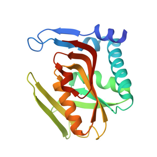 | |
UniProt & NIH Common Fund Data Resources | |||||
Find proteins for P07741 (Homo sapiens) Explore P07741 Go to UniProtKB: P07741 | |||||
PHAROS: P07741 GTEx: ENSG00000198931 | |||||
Entity Groups | |||||
| Sequence Clusters | 30% Identity50% Identity70% Identity90% Identity95% Identity100% Identity | ||||
| UniProt Group | P07741 | ||||
Sequence AnnotationsExpand | |||||
| |||||
| Ligands 3 Unique | |||||
|---|---|---|---|---|---|
| ID | Chains | Name / Formula / InChI Key | 2D Diagram | 3D Interactions | |
| AMP Query on AMP | B [auth A] | ADENOSINE MONOPHOSPHATE C10 H14 N5 O7 P UDMBCSSLTHHNCD-KQYNXXCUSA-N |  | ||
| SO4 Query on SO4 | C [auth A] | SULFATE ION O4 S QAOWNCQODCNURD-UHFFFAOYSA-L |  | ||
| GOL Query on GOL | D [auth A] | GLYCEROL C3 H8 O3 PEDCQBHIVMGVHV-UHFFFAOYSA-N |  | ||
| Length ( Å ) | Angle ( ˚ ) |
|---|---|
| a = 59.493 | α = 90 |
| b = 59.493 | β = 90 |
| c = 109.772 | γ = 90 |
| Software Name | Purpose |
|---|---|
| MOSFLM | data reduction |
| SCALA | data scaling |
| PHENIX | refinement |
| PDB_EXTRACT | data extraction |
| MOLREP | phasing |
RCSB PDB (citation) is hosted by
RCSB PDB is a member of the

