4X3T

Crystal structure of chromobox homolog 7 (CBX7) chromodomain with MS37452


Experimental Data Snapshot

  • Method: X-RAY DIFFRACTION
  • Resolution: 2.14 Å
  • R-Value Free: 0.256 
  • R-Value Work: 0.203 
  • R-Value Observed: 0.206 

wwPDB Validation   3D Report Full Report


Ligand Structure Quality Assessment 


This is version 1.1 of the entry. See complete history


Literature

Small-Molecule Modulators of Methyl-Lysine Binding for the CBX7 Chromodomain.

Ren, C.Morohashi, K.Plotnikov, A.N.Jakoncic, J.Smith, S.G.Li, J.Zeng, L.Rodriguez, Y.Stojanoff, V.Walsh, M.Zhou, M.M.

(2015) Chem Biol 22: 161-168

  • DOI: https://doi.org/10.1016/j.chembiol.2014.11.021
  • Primary Citation of Related Structures:  
    4X3K, 4X3S, 4X3T, 4X3U

  • PubMed Abstract: 

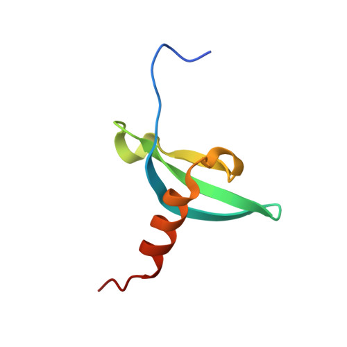
    Chromobox homolog 7 (CBX7) plays an important role in gene transcription in a wide array of cellular processes, ranging from stem cell self-renewal and differentiation to tumor progression. CBX7 functions through its N-terminal chromodomain (ChD), which recognizes trimethylated lysine 27 of histone 3 (H3K27me3), a conserved epigenetic mark that signifies gene transcriptional repression. In this study, we report the discovery of small molecules that inhibit CBX7ChD binding to H3K27me3. Our crystal structures reveal the binding modes of these molecules that compete against H3K27me3 binding through interactions with key residues in the methyl-lysine binding pocket of CBX7ChD. We further show that a lead compound, MS37452, derepresses transcription of Polycomb repressive complex target gene p16/CDKN2A by displacing CBX7 binding to the INK4A/ARF locus in prostate cancer cells. These small molecules have the potential to be developed into high-potency chemical modulators that target CBX7 functions in gene transcription in different disease pathways.


  • Organizational Affiliation

    Department of Structural and Chemical Biology, Icahn School of Medicine at Mount Sinai, 1425 Madison Avenue, New York, NY 10029, USA.


Macromolecules
Find similar proteins by:  (by identity cutoff)  |  3D Structure
Entity ID: 1
MoleculeChains Sequence LengthOrganismDetailsImage
Chromobox protein homolog 7
A, B, C, D, E
A, B, C, D, E, F
64Mus musculusMutation(s): 0 
Gene Names: Cbx7D15Ertd417e
UniProt
Find proteins for Q8VDS3 (Mus musculus)
Explore Q8VDS3 
Go to UniProtKB:  Q8VDS3
Entity Groups  
Sequence Clusters30% Identity50% Identity70% Identity90% Identity95% Identity100% Identity
UniProt GroupQ8VDS3
Sequence Annotations
Expand
  • Reference Sequence
Small Molecules
Ligands 3 Unique
IDChains Name / Formula / InChI Key2D Diagram3D Interactions
45E
Query on 45E

Download Ideal Coordinates CCD File 
G [auth A]
J [auth B]
L [auth C]
O [auth D]
R [auth E]
G [auth A],
J [auth B],
L [auth C],
O [auth D],
R [auth E],
T [auth F]
1-[4-(2,3-dimethoxybenzoyl)piperazin-1-yl]-2-(3-methylphenoxy)ethanone
C22 H26 N2 O5
LUMCNRKHZRYQOV-UHFFFAOYSA-N
ZN
Query on ZN

Download Ideal Coordinates CCD File 
H [auth A]
I [auth A]
K [auth B]
M [auth C]
N [auth C]
H [auth A],
I [auth A],
K [auth B],
M [auth C],
N [auth C],
P [auth D],
Q [auth D],
S [auth E],
U [auth F]
ZINC ION
Zn
PTFCDOFLOPIGGS-UHFFFAOYSA-N
EDO
Query on EDO

Download Ideal Coordinates CCD File 
V [auth F]1,2-ETHANEDIOL
C2 H6 O2
LYCAIKOWRPUZTN-UHFFFAOYSA-N
Binding Affinity Annotations 
IDSourceBinding Affinity
45E BindingDB:  4X3T Ki: 3.31e+4 (nM) from 1 assay(s)
Kd: min: 2.90e+4, max: 1.25e+5 (nM) from 2 assay(s)
IC50: 6.70e+4 (nM) from 1 assay(s)
Binding MOAD:  4X3T Kd: 2.89e+4 (nM) from 1 assay(s)
Experimental Data & Validation

Experimental Data

  • Method: X-RAY DIFFRACTION
  • Resolution: 2.14 Å
  • R-Value Free: 0.256 
  • R-Value Work: 0.203 
  • R-Value Observed: 0.206 
  • Space Group: P 1 21 1
Unit Cell:
Length ( Å )Angle ( ˚ )
a = 54.004α = 90
b = 77.309β = 95.55
c = 66.842γ = 90
Software Package:
Software NamePurpose
REFMACrefinement
HKL-2000data reduction
HKL-2000data scaling
PHENIXphasing

Structure Validation

View Full Validation Report


Ligand Structure Quality Assessment 


Entry History 

Deposition Data

Revision History  (Full details and data files)

  • Version 1.0: 2015-03-04
    Type: Initial release
  • Version 1.1: 2024-02-28
    Changes: Data collection, Database references, Derived calculations, Source and taxonomy