4X3J

Selection of fragments for kinase inhibitor design: decoration is key


Experimental Data Snapshot

  • Method: X-RAY DIFFRACTION
  • Resolution: 2.50 Å
  • R-Value Free: 0.293 
  • R-Value Work: 0.255 
  • R-Value Observed: 0.257 

wwPDB Validation   3D Report Full Report


Ligand Structure Quality Assessment 


This is version 1.1 of the entry. See complete history


Literature

Selection of fragments for kinase inhibitor design: decoration is key.

Czodrowski, P.Holzemann, G.Barnickel, G.Greiner, H.Musil, D.

(2015) J Med Chem 58: 457-465

  • DOI: https://doi.org/10.1021/jm501597j
  • Primary Citation of Related Structures:  
    4X0M, 4X2F, 4X2G, 4X2J, 4X2K, 4X2N, 4X3J

  • PubMed Abstract: 

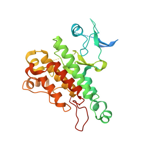
    In fragment-based screening, the choice of the best suited fragment hit among the detected hits is crucial for success. In our study, a kinase lead compound was fragmented, the hinge-binding motif extracted as a core fragment, and a minilibrary of five similar compounds with fragment-like properties was selected from our proprietary compound database. The structures of five fragments in complex with transforming growth factor β receptor type 1 kinase domain were determined by X-ray crystallography. Three different binding modes of the fragments are observed that depend on the position and the type of the substitution at the core fragment. The influence of different substituents on the preferred fragment pose was analyzed by various computational approaches. We postulate that the replacement of water molecules leads to the different binding modes.


  • Organizational Affiliation

    Discovery Technologies, Merck Serono Research, Merck Serono R&D, Merck KGaA , Frankfurter Strasse 250, 64293 Darmstadt, Germany.


Macromolecules
Find similar proteins by:  (by identity cutoff)  |  3D Structure
Entity ID: 1
MoleculeChains Sequence LengthOrganismDetailsImage
Angiopoietin-1 receptor321Homo sapiensMutation(s): 1 
Gene Names: TEKTIE2VMCMVMCM1
EC: 2.7.10.1
UniProt & NIH Common Fund Data Resources
Find proteins for Q02763 (Homo sapiens)
Explore Q02763 
Go to UniProtKB:  Q02763
PHAROS:  Q02763
GTEx:  ENSG00000120156 
Entity Groups  
Sequence Clusters30% Identity50% Identity70% Identity90% Identity95% Identity100% Identity
UniProt GroupQ02763
Sequence Annotations
Expand
  • Reference Sequence
Small Molecules
Ligands 1 Unique
IDChains Name / Formula / InChI Key2D Diagram3D Interactions
3WR
Query on 3WR

Download Ideal Coordinates CCD File 
B [auth A]1-[4-(4-amino-5-oxopyrido[2,3-d]pyrimidin-8(5H)-yl)phenyl]-3-[2-fluoro-5-(trifluoromethyl)phenyl]urea
C21 H14 F4 N6 O2
OWKPAZCWJGPSQU-UHFFFAOYSA-N
Experimental Data & Validation

Experimental Data

  • Method: X-RAY DIFFRACTION
  • Resolution: 2.50 Å
  • R-Value Free: 0.293 
  • R-Value Work: 0.255 
  • R-Value Observed: 0.257 
  • Space Group: P 41 21 2
Unit Cell:
Length ( Å )Angle ( ˚ )
a = 62.456α = 90
b = 62.456β = 90
c = 178.45γ = 90
Software Package:
Software NamePurpose
BUSTERrefinement
DENZOdata reduction
SCALEPACKdata scaling

Structure Validation

View Full Validation Report


Ligand Structure Quality Assessment 


Entry History 

Deposition Data

Revision History  (Full details and data files)

  • Version 1.0: 2014-12-24
    Type: Initial release
  • Version 1.1: 2015-01-21
    Changes: Database references