4WZS

Crystal structure of the Mot1 N-terminal domain in complex with TBP and NC2 bound to a promoter DNA fragment


Experimental Data Snapshot

  • Method: X-RAY DIFFRACTION
  • Resolution: 3.78 Å
  • R-Value Free: 0.258 
  • R-Value Work: 0.235 
  • R-Value Observed: 0.236 

wwPDB Validation   3D Report Full Report


This is version 1.2 of the entry. See complete history


Literature

Structural basis for recognition and remodeling of the TBP:DNA:NC2 complex by Mot1.

Butryn, A.Schuller, J.M.Stoehr, G.Runge-Wollmann, P.Forster, F.Auble, D.T.Hopfner, K.P.

(2015) Elife 4

  • DOI: https://doi.org/10.7554/eLife.07432
  • Primary Citation of Related Structures:  
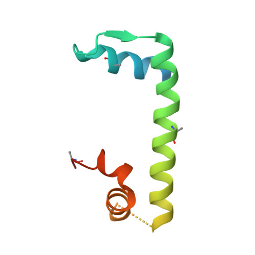
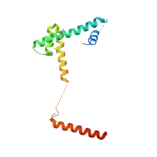
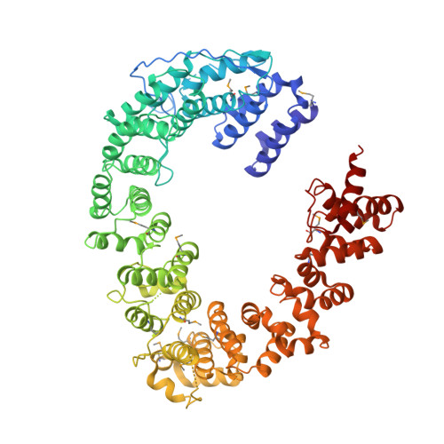
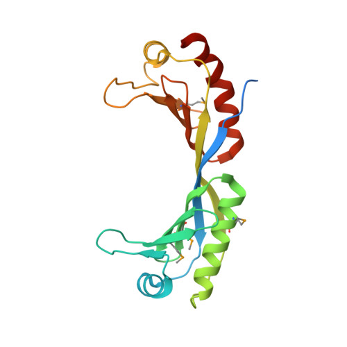
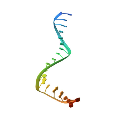
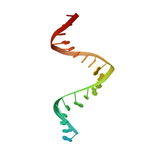
    4WZS

  • PubMed Abstract: 

    Swi2/Snf2 ATPases remodel substrates such as nucleosomes and transcription complexes to control a wide range of DNA-associated processes, but detailed structural information on the ATP-dependent remodeling reactions is largely absent. The single subunit remodeler Mot1 (modifier of transcription 1) dissociates TATA box-binding protein (TBP):DNA complexes, offering a useful system to address the structural mechanisms of Swi2/Snf2 ATPases. Here, we report the crystal structure of the N-terminal domain of Mot1 in complex with TBP, DNA, and the transcription regulator negative cofactor 2 (NC2). Our data show that Mot1 reduces DNA:NC2 interactions and unbends DNA as compared to the TBP:DNA:NC2 state, suggesting that Mot1 primes TBP:NC2 displacement in an ATP-independent manner. Electron microscopy and cross-linking data suggest that the Swi2/Snf2 domain of Mot1 associates with the upstream DNA and the histone fold of NC2, thereby revealing parallels to some nucleosome remodelers. This study provides a structural framework for how a Swi2/Snf2 ATPase interacts with its substrate DNA:protein complex.


  • Organizational Affiliation

    Gene Center, Department of Biochemistry, Ludwig Maximilian University, Munich, Germany.


Macromolecules

Find similar proteins by:  (by identity cutoff)  |  3D Structure
Entity ID: 1
MoleculeChains Sequence LengthOrganismDetailsImage
ECU11_1470 protein95Encephalitozoon cuniculi GB-M1Mutation(s): 0 
Gene Names: ECU11_1470
UniProt
Find proteins for Q8SQT6 (Encephalitozoon cuniculi (strain GB-M1))
Explore Q8SQT6 
Go to UniProtKB:  Q8SQT6
Entity Groups  
Sequence Clusters30% Identity50% Identity70% Identity90% Identity95% Identity100% Identity
UniProt GroupQ8SQT6
Sequence Annotations
Expand
  • Reference Sequence
Find similar proteins by:  (by identity cutoff)  |  3D Structure
Entity ID: 2
MoleculeChains Sequence LengthOrganismDetailsImage
TATA-binding protein-associated phosphoprotein149Encephalitozoon cuniculiMutation(s): 0 
Gene Names: ECU11_0730
UniProt
Find proteins for M1K2J7 (Encephalitozoon cuniculi)
Explore M1K2J7 
Go to UniProtKB:  M1K2J7
Entity Groups  
Sequence Clusters30% Identity50% Identity70% Identity90% Identity95% Identity100% Identity
UniProt GroupM1K2J7
Sequence Annotations
Expand
  • Reference Sequence
Find similar proteins by:  (by identity cutoff)  |  3D Structure
Entity ID: 3
MoleculeChains Sequence LengthOrganismDetailsImage
Similarity to HELICASE MOT1780Encephalitozoon cuniculi GB-M1Mutation(s): 0 
Gene Names: ECU03_1530
UniProt
Find proteins for Q8SVZ5 (Encephalitozoon cuniculi (strain GB-M1))
Explore Q8SVZ5 
Go to UniProtKB:  Q8SVZ5
Entity Groups  
Sequence Clusters30% Identity50% Identity70% Identity90% Identity95% Identity100% Identity
UniProt GroupQ8SVZ5
Sequence Annotations
Expand
  • Reference Sequence
Find similar proteins by:  (by identity cutoff)  |  3D Structure
Entity ID: 4
MoleculeChains Sequence LengthOrganismDetailsImage
ECU04_1440 protein200Encephalitozoon cuniculi GB-M1Mutation(s): 0 
Gene Names: ECU04_1440
UniProt
Find proteins for Q8ST28 (Encephalitozoon cuniculi (strain GB-M1))
Explore Q8ST28 
Go to UniProtKB:  Q8ST28
Entity Groups  
Sequence Clusters30% Identity50% Identity70% Identity90% Identity95% Identity100% Identity
UniProt GroupQ8ST28
Sequence Annotations
Expand
  • Reference Sequence

Find similar nucleic acids by:  Sequence   |   3D Structure  

Entity ID: 5
MoleculeChains LengthOrganismImage
DNA (5'-D(P*CP*CP*AP*CP*CP*CP*CP*CP*TP*TP*TP*TP*AP*TP*AP*GP*CP*CP*CP*T)-3')24synthetic construct
Sequence Annotations
Expand
  • Reference Sequence

Find similar nucleic acids by:  Sequence   |   3D Structure  

Entity ID: 6
MoleculeChains LengthOrganismImage
DNA (5'-D(P*GP*GP*GP*CP*TP*AP*TP*AP*AP*AP*AP*GP*GP*GP*GP*GP*TP*GP*G)-3')24synthetic construct
Sequence Annotations
Expand
  • Reference Sequence
Small Molecules
Modified Residues  1 Unique
IDChains TypeFormula2D DiagramParent
MSE
Query on MSE
A
L-PEPTIDE LINKINGC5 H11 N O2 SeMET
Experimental Data & Validation

Experimental Data

  • Method: X-RAY DIFFRACTION
  • Resolution: 3.78 Å
  • R-Value Free: 0.258 
  • R-Value Work: 0.235 
  • R-Value Observed: 0.236 
  • Space Group: C 1 2 1
Unit Cell:
Length ( Å )Angle ( ˚ )
a = 150.629α = 90
b = 140.274β = 113.7
c = 90.778γ = 90
Software Package:
Software NamePurpose
BUSTERrefinement
PHASERphasing
XDSdata reduction
XSCALEdata scaling

Structure Validation

View Full Validation Report


Entry History & Funding Information

Deposition Data


Funding OrganizationLocationGrant Number
German Research FoundationGermanySFB 646

Revision History  (Full details and data files)

  • Version 1.0: 2015-08-26
    Type: Initial release
  • Version 1.1: 2017-09-06
    Changes: Author supporting evidence, Data collection, Derived calculations
  • Version 1.2: 2024-01-10
    Changes: Data collection, Database references, Refinement description