4WZ5

Crystal structure of P. aeruginosa OXA10


Experimental Data Snapshot

  • Method: X-RAY DIFFRACTION
  • Resolution: 1.60 Å
  • R-Value Free: 0.208 
  • R-Value Work: 0.190 
  • R-Value Observed: 0.191 

wwPDB Validation   3D Report Full Report


Ligand Structure Quality Assessment 


This is version 1.1 of the entry. See complete history


Literature

4,5-Disubstituted 6-Aryloxy-1,3-dihydrobenzo[c][1,2]oxaboroles Are Broad-Spectrum Serine beta-Lactamase Inhibitors.

McKinney, D.C.Zhou, F.Eyermann, C.J.Ferguson, A.D.Prince, D.B.Breen, J.Giacobbe, R.A.Lahiri, S.Verheijen, J.C.

(2015) ACS Infect Dis 1: 310-316

  • DOI: https://doi.org/10.1021/acsinfecdis.5b00031
  • Primary Citation of Related Structures:  
    4WYY, 4WZ4, 4WZ5

  • PubMed Abstract: 

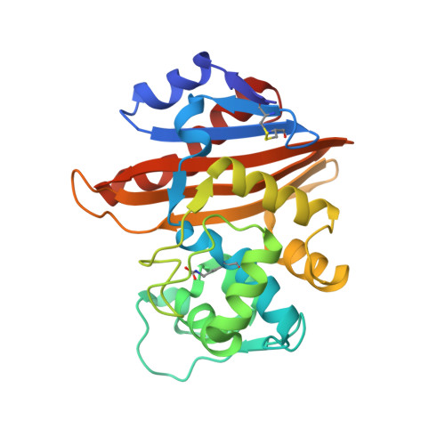
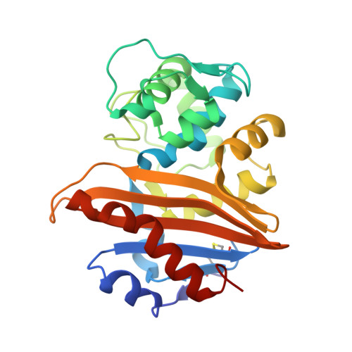
    Bacterially expressed β-lactamases are rapidly eroding the clinical utility of the important β-lactam class of antibacterials, significantly impairing our ability to fight serious bacterial infections. This paper describes a study of oxaborole-derived β-lactamase inhibitors in which crystal structures and computational modeling aided in the rational design of analogues with improved spectrum of activity against class A, C, and D enzymes. Crystal structures of two of these inhibitors covalently bound to two different serine β-lactamases, class C Pseudomonas aeruginosa AmpC and class D OXA-10, are described herein. Improved physicochemical properties as well as increased activity against an array of β-lactamases resulted in substantial restoration of susceptibility to ceftazidime in Escherichia coli and Klebsiella pneumoniae.


  • Organizational Affiliation

    Infection Innovative Medicines, AstraZeneca R&D Boston , 35 Gatehouse Drive, Waltham Massachusetts 02451, United States.


Macromolecules
Find similar proteins by:  (by identity cutoff)  |  3D Structure
Entity ID: 1
MoleculeChains Sequence LengthOrganismDetailsImage
Beta-lactamase OXA-10
A, B
248Pseudomonas aeruginosaMutation(s): 0 
Gene Names: blaoxa10pse2
EC: 3.5.2.6
UniProt
Find proteins for P14489 (Pseudomonas aeruginosa)
Explore P14489 
Go to UniProtKB:  P14489
Entity Groups  
Sequence Clusters30% Identity50% Identity70% Identity90% Identity95% Identity100% Identity
UniProt GroupP14489
Sequence Annotations
Expand
  • Reference Sequence
Find similar proteins by:  (by identity cutoff)  |  3D Structure
Entity ID: 2
MoleculeChains Sequence LengthOrganismDetailsImage
Beta-lactamase OXA-10
C, D
248Pseudomonas aeruginosaMutation(s): 0 
Gene Names: blaoxa10pse2
EC: 3.5.2.6
UniProt
Find proteins for P14489 (Pseudomonas aeruginosa)
Explore P14489 
Go to UniProtKB:  P14489
Entity Groups  
Sequence Clusters30% Identity50% Identity70% Identity90% Identity95% Identity100% Identity
UniProt GroupP14489
Sequence Annotations
Expand
  • Reference Sequence
Small Molecules
Ligands 3 Unique
IDChains Name / Formula / InChI Key2D Diagram3D Interactions
3VU
Query on 3VU

Download Ideal Coordinates CCD File 
G [auth A],
K [auth B]
{(3R)-6-[(3-amino-1,2,4-thiadiazol-5-yl)oxy]-1-hydroxy-4,5-dimethyl-1,3-dihydro-2,1-benzoxaborol-3-yl}acetic acid
C13 H14 B N3 O5 S
CXFHZAGALDYYDH-SECBINFHSA-N
SO4
Query on SO4

Download Ideal Coordinates CCD File 
E [auth A]
F [auth A]
H [auth B]
I [auth B]
J [auth B]
E [auth A],
F [auth A],
H [auth B],
I [auth B],
J [auth B],
L [auth C],
M [auth C],
N [auth C],
O [auth C],
Q [auth D],
R [auth D],
S [auth D],
T [auth D]
SULFATE ION
O4 S
QAOWNCQODCNURD-UHFFFAOYSA-L
CO2
Query on CO2

Download Ideal Coordinates CCD File 
P [auth C],
U [auth D]
CARBON DIOXIDE
C O2
CURLTUGMZLYLDI-UHFFFAOYSA-N
Modified Residues  1 Unique
IDChains TypeFormula2D DiagramParent
KCX
Query on KCX
A, B
L-PEPTIDE LINKINGC7 H14 N2 O4LYS
Experimental Data & Validation

Experimental Data

  • Method: X-RAY DIFFRACTION
  • Resolution: 1.60 Å
  • R-Value Free: 0.208 
  • R-Value Work: 0.190 
  • R-Value Observed: 0.191 
  • Space Group: P 1 21 1
Unit Cell:
Length ( Å )Angle ( ˚ )
a = 65.74α = 90
b = 82.35β = 94.54
c = 102.38γ = 90
Software Package:
Software NamePurpose
BUSTERrefinement
XDSdata reduction
Aimlessdata scaling
MOLREPphasing
Cootmodel building

Structure Validation

View Full Validation Report


Ligand Structure Quality Assessment 


Entry History 

Deposition Data

Revision History  (Full details and data files)

  • Version 1.0: 2015-08-05
    Type: Initial release
  • Version 1.1: 2016-09-28
    Changes: Database references