4WY7

Crystal structure of recombinant 4E10 expressed in Escherichia coli with epitope bound


Experimental Data Snapshot

  • Method: X-RAY DIFFRACTION
  • Resolution: 2.10 Å
  • R-Value Free: 0.207 
  • R-Value Work: 0.162 
  • R-Value Observed: 0.164 

wwPDB Validation   3D Report Full Report


This is version 1.3 of the entry. See complete history


Literature

The Atomic Structure of the HIV-1 gp41 Transmembrane Domain and Its Connection to the Immunogenic Membrane-proximal External Region.

Apellaniz, B.Rujas, E.Serrano, S.Morante, K.Tsumoto, K.Caaveiro, J.M.Jimenez, M.A.Nieva, J.L.

(2015) J Biol Chem 290: 12999-13015

  • DOI: https://doi.org/10.1074/jbc.M115.644351
  • Primary Citation of Related Structures:  
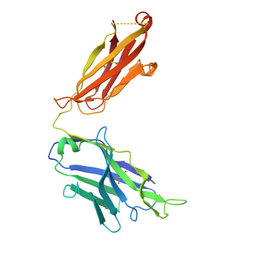
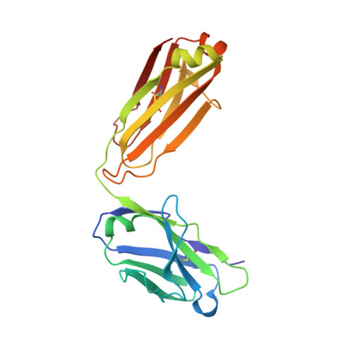
    2MG1, 2MG2, 2MG3, 4WY7

  • PubMed Abstract: 

    The membrane-proximal external region (MPER) C-terminal segment and the transmembrane domain (TMD) of gp41 are involved in HIV-1 envelope glycoprotein-mediated fusion and modulation of immune responses during viral infection. However, the atomic structure of this functional region remains unsolved. Here, based on the high resolution NMR data obtained for peptides spanning the C-terminal segment of MPER and the TMD, we report two main findings: (i) the conformational variability of the TMD helix at a membrane-buried position; and (ii) the existence of an uninterrupted α-helix spanning MPER and the N-terminal region of the TMD. Thus, our structural data provide evidence for the bipartite organization of TMD predicted by previous molecular dynamics simulations and functional studies, but they do not support the breaking of the helix at Lys-683, as was suggested by some models to mark the initiation of the TMD anchor. Antibody binding energetics examined with isothermal titration calorimetry and humoral responses elicited in rabbits by peptide-based vaccines further support the relevance of a continuous MPER-TMD helix for immune recognition. We conclude that the transmembrane anchor of HIV-1 envelope is composed of two distinct subdomains: 1) an immunogenic helix at the N terminus also involved in promoting membrane fusion; and 2) an immunosuppressive helix at the C terminus, which might also contribute to the late stages of the fusion process. The unprecedented high resolution structural data reported here may guide future vaccine and inhibitor developments.


  • Organizational Affiliation

    From the Biophysics Unit (Consejo Superior de Investigaciones Científicas, UPV/EHU) and Department of Biochemistry and Molecular Biology, University of the Basque Country (UPV/EHU), P. O. Box 644, 48080 Bilbao, Spain.


Macromolecules
Find similar proteins by:  (by identity cutoff)  |  3D Structure
Entity ID: 1
MoleculeChains Sequence LengthOrganismDetailsImage
Fab 4E10 Heavy chainA [auth H]228Homo sapiensMutation(s): 0 
Entity Groups  
Sequence Clusters30% Identity50% Identity70% Identity90% Identity95% Identity100% Identity
Sequence Annotations
Expand
  • Reference Sequence
Find similar proteins by:  (by identity cutoff)  |  3D Structure
Entity ID: 2
MoleculeChains Sequence LengthOrganismDetailsImage
Fab 4E10Light chainB [auth L]214Homo sapiensMutation(s): 0 
Entity Groups  
Sequence Clusters30% Identity50% Identity70% Identity90% Identity95% Identity100% Identity
Sequence Annotations
Expand
  • Reference Sequence

Find similar proteins by:  Sequence   |   3D Structure  

Entity ID: 3
MoleculeChains Sequence LengthOrganismDetailsImage
Envelope glycoprotein gp160C [auth P]16Human immunodeficiency virus type 1 (WMJ2 ISOLATE)Mutation(s): 0 
Gene Names: env
UniProt
Find proteins for P05880 (Human immunodeficiency virus type 1 group M subtype B (isolate WMJ22))
Explore P05880 
Go to UniProtKB:  P05880
Entity Groups  
Sequence Clusters30% Identity50% Identity70% Identity90% Identity95% Identity100% Identity
UniProt GroupP05880
Sequence Annotations
Expand
  • Reference Sequence
Experimental Data & Validation

Experimental Data

  • Method: X-RAY DIFFRACTION
  • Resolution: 2.10 Å
  • R-Value Free: 0.207 
  • R-Value Work: 0.162 
  • R-Value Observed: 0.164 
  • Space Group: C 1 2 1
Unit Cell:
Length ( Å )Angle ( ˚ )
a = 157.3α = 90
b = 44.58β = 113.8
c = 85.4γ = 90
Software Package:
Software NamePurpose
REFMACrefinement
MOSFLMdata reduction
SCALAdata scaling
PHASERphasing
Cootmodel building

Structure Validation

View Full Validation Report


Entry History 

Deposition Data

Revision History  (Full details and data files)

  • Version 1.0: 2015-03-25
    Type: Initial release
  • Version 1.1: 2015-04-01
    Changes: Database references
  • Version 1.2: 2015-06-03
    Changes: Database references
  • Version 1.3: 2024-01-10
    Changes: Data collection, Database references, Refinement description