4WW3

Crystal structure of the lumi intermediate of squid rhodopsin


Experimental Data Snapshot

  • Method: X-RAY DIFFRACTION
  • Resolution: 2.80 Å
  • R-Value Free: 0.296 
  • R-Value Work: 0.290 
  • R-Value Observed: 0.290 

wwPDB Validation   3D Report Full Report


Ligand Structure Quality Assessment 


This is version 1.3 of the entry. See complete history


Literature

Crystallographic Study of the LUMI Intermediate of Squid Rhodopsin

Murakami, M.Kouyama, T.

(2015) PLoS One 10: e0126970-e0126970

  • DOI: https://doi.org/10.1371/journal.pone.0126970
  • Primary Citation of Related Structures:  
    4WW3

  • PubMed Abstract: 

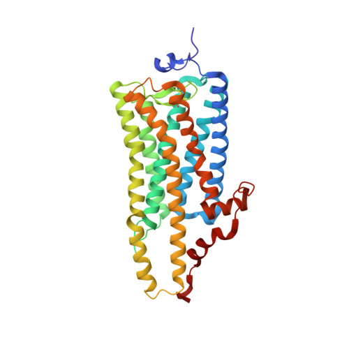
    Upon absorption of light, the retinal chromophore in rhodopsin isomerizes from the 11-cis to the trans configuration, initiating a photoreaction cycle. The primary photoreaction state, bathorhodopsin (BATHO), relaxes thermally through lumirhodopsin (LUMI) into a photoactive state, metarhodopsin (META), which stimulates the conjugated G-protein. Previous crystallographic studies of squid and bovine rhodopsins have shown that the structural change in the primary photoreaction of squid rhodopsin is considerably different from that observed in bovine rhodopsin. It would be expected that there is a fundamental difference in the subsequent thermal relaxation process between vertebrate and invertebrate rhodopsins. In this work, we performed crystallographic analyses of the LUMI state of squid rhodopsin using the P62 crystal. When the crystal was illuminated at 100 K with blue light, a half fraction of the protein was converted into BATHO. This reaction state relaxed into LUMI when the illuminated crystal was warmed in the dark to 170 K. It was found that, whereas trans retinal is largely twisted in BATHO, it takes on a more planar configuration in LUMI. This relaxation of retinal is accompanied by reorientation of the Schiff base NH bond, the hydrogen-bonding partner of which is switched to Asn185 in LUMI. Unlike bovine rhodopsin, the BATHO-to-LUMI transition in squid rhodopsin was accompanied by no significant change in the position/orientation of the beta-ionone ring of retinal.


  • Organizational Affiliation

    Department of Physics, Graduate School of Science, Nagoya University, Nagoya, Japan.


Macromolecules
Find similar proteins by:  (by identity cutoff)  |  3D Structure
Entity ID: 1
MoleculeChains Sequence LengthOrganismDetailsImage
Rhodopsin
A, B
350Todarodes pacificusMutation(s): 0 
Membrane Entity: Yes 
UniProt
Find proteins for P31356 (Todarodes pacificus)
Explore P31356 
Go to UniProtKB:  P31356
Entity Groups  
Sequence Clusters30% Identity50% Identity70% Identity90% Identity95% Identity100% Identity
UniProt GroupP31356
Sequence Annotations
Expand
  • Reference Sequence
Small Molecules
Ligands 6 Unique
IDChains Name / Formula / InChI Key2D Diagram3D Interactions
PC1
Query on PC1

Download Ideal Coordinates CCD File 
M [auth B]1,2-DIACYL-SN-GLYCERO-3-PHOSPHOCHOLINE
C44 H88 N O8 P
NRJAVPSFFCBXDT-HUESYALOSA-N
TWT
Query on TWT

Download Ideal Coordinates CCD File 
F [auth A],
L [auth B]
DOCOSANE
C22 H46
HOWGUJZVBDQJKV-UHFFFAOYSA-N
BOG
Query on BOG

Download Ideal Coordinates CCD File 
G [auth A],
N [auth B]
octyl beta-D-glucopyranoside
C14 H28 O6
HEGSGKPQLMEBJL-RKQHYHRCSA-N
RET
Query on RET

Download Ideal Coordinates CCD File 
C [auth A],
I [auth B]
RETINAL
C20 H28 O
NCYCYZXNIZJOKI-OVSJKPMPSA-N
PLM
Query on PLM

Download Ideal Coordinates CCD File 
D [auth A],
E [auth A],
J [auth B],
K [auth B]
PALMITIC ACID
C16 H32 O2
IPCSVZSSVZVIGE-UHFFFAOYSA-N
SO4
Query on SO4

Download Ideal Coordinates CCD File 
H [auth B]SULFATE ION
O4 S
QAOWNCQODCNURD-UHFFFAOYSA-L
Experimental Data & Validation

Experimental Data

  • Method: X-RAY DIFFRACTION
  • Resolution: 2.80 Å
  • R-Value Free: 0.296 
  • R-Value Work: 0.290 
  • R-Value Observed: 0.290 
  • Space Group: P 62
Unit Cell:
Length ( Å )Angle ( ˚ )
a = 122.39α = 90
b = 122.39β = 90
c = 158.77γ = 120
Software Package:
Software NamePurpose
CNSrefinement
MOSFLMdata reduction
SCALAdata scaling
CNSphasing
PDB_EXTRACTdata extraction

Structure Validation

View Full Validation Report


Ligand Structure Quality Assessment 


Entry History & Funding Information

Deposition Data


Funding OrganizationLocationGrant Number
Ministry of Education, Science, Sports and Culture(MEXT)Japan23121513
Ministry of Education, Science, Sports and Culture(MEXT)Japan24370043

Revision History  (Full details and data files)

  • Version 1.0: 2015-06-17
    Type: Initial release
  • Version 1.1: 2020-02-05
    Changes: Data collection, Derived calculations
  • Version 1.2: 2020-07-29
    Type: Remediation
    Reason: Carbohydrate remediation
    Changes: Data collection, Derived calculations, Structure summary
  • Version 1.3: 2023-11-08
    Changes: Data collection, Database references, Refinement description, Structure summary