4WVP

Crystal structure of an activity-based probe HNE complex


Experimental Data Snapshot

  • Method: X-RAY DIFFRACTION
  • Resolution: 1.63 Å
  • R-Value Free: 0.169 
  • R-Value Work: 0.138 
  • R-Value Observed: 0.140 

wwPDB Validation   3D Report Full Report


This is version 3.0 of the entry. See complete history


Literature

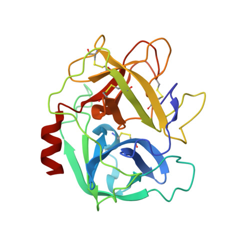
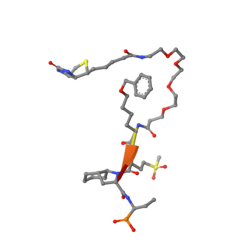
The Elastase-PK101 Structure: Mechanism of an Ultrasensitive Activity-based Probe Revealed.

Lechtenberg, B.C.Kasperkiewicz, P.Robinson, H.Drag, M.Riedl, S.J.

(2015) ACS Chem Biol 10: 945-951

  • DOI: https://doi.org/10.1021/cb500909n
  • Primary Citation of Related Structures:  
    4WVP

  • PubMed Abstract: 

    Human neutrophil elastase (HNE) plays a central role in neutrophil host defense, but its broad specificity makes HNE a difficult target for both inhibitor and probe development. Recently, we identified the unnatural amino acid containing activity-based probe PK101, which exhibits astounding sensitivity and selectivity for HNE, yet completely lacks mechanistic explanation for its unique characteristics. Here, we present the crystal structure of the HNE-PK101 complex which not only reveals the basis for PK101 ultrasensitivity but also uncovers so far unrecognized HNE features. Strikingly, the Nle(O-Bzl) function in the P4 position of PK101 reveals and leverages an "exo-pocket" on HNE as a critical factor for selectivity. Furthermore, the PK101 P3 position harbors a methionine dioxide function, which mimics a post-translationally oxidized methionine residue and forms a critical hydrogen bond to the backbone amide of Gly219 of HNE. Gly219 resides in a Gly-Gly motif that is unique to HNE, yet compulsory for this interaction. Consequently, this feature enables HNE to accommodate substrates that have undergone methionine oxidation, which constitutes a hallmark post-translational modification of neutrophil signaling.


  • Organizational Affiliation

    †Sanford-Burnham Medical Research Institute, 10901 North Torrey Pines Road, La Jolla, California 92037, United States.


Macromolecules
Find similar proteins by:  (by identity cutoff)  |  3D Structure
Entity ID: 1
MoleculeChains Sequence LengthOrganismDetailsImage
Neutrophil elastaseA [auth E]218Homo sapiensMutation(s): 0 
EC: 3.4.21.37
UniProt & NIH Common Fund Data Resources
Find proteins for P08246 (Homo sapiens)
Explore P08246 
Go to UniProtKB:  P08246
PHAROS:  P08246
GTEx:  ENSG00000197561 
Entity Groups  
Sequence Clusters30% Identity50% Identity70% Identity90% Identity95% Identity100% Identity
UniProt GroupP08246
Sequence Annotations
Expand
  • Reference Sequence

Find similar proteins by:  Sequence   |   3D Structure  

Entity ID: 2
MoleculeChains Sequence LengthOrganismDetailsImage
BTN-3V3-NLB-OMT-OIC-3V2B [auth I]6synthetic constructMutation(s): 0 
Sequence Annotations
Expand
  • Reference Sequence
Oligosaccharides

Help

Entity ID: 3
MoleculeChains Length2D Diagram Glycosylation3D Interactions
beta-D-mannopyranose-(1-4)-2-acetamido-2-deoxy-beta-D-glucopyranose-(1-4)-[alpha-L-fucopyranose-(1-6)]2-acetamido-2-deoxy-beta-D-glucopyranoseC [auth A]4N-Glycosylation
Glycosylation Resources
GlyTouCan:  G32152BH
GlyCosmos:  G32152BH
GlyGen:  G32152BH
Entity ID: 4
MoleculeChains Length2D Diagram Glycosylation3D Interactions
2-acetamido-2-deoxy-beta-D-glucopyranose-(1-4)-[alpha-L-fucopyranose-(1-6)]2-acetamido-2-deoxy-beta-D-glucopyranoseD [auth B]3N-Glycosylation
Glycosylation Resources
GlyTouCan:  G21290RB
GlyCosmos:  G21290RB
GlyGen:  G21290RB
Small Molecules
Modified Residues  2 Unique
IDChains TypeFormula2D DiagramParent
NLB
Query on NLB
B [auth I]L-PEPTIDE LINKINGC13 H19 N O3LEU
OMT
Query on OMT
B [auth I]L-PEPTIDE LINKINGC5 H11 N O4 SMET
Biologically Interesting Molecules (External Reference) 1 Unique
Experimental Data & Validation

Experimental Data

  • Method: X-RAY DIFFRACTION
  • Resolution: 1.63 Å
  • R-Value Free: 0.169 
  • R-Value Work: 0.138 
  • R-Value Observed: 0.140 
  • Space Group: P 63
Unit Cell:
Length ( Å )Angle ( ˚ )
a = 73.12α = 90
b = 73.12β = 90
c = 69.451γ = 120
Software Package:
Software NamePurpose
MOSFLMdata reduction
Aimlessdata scaling
PHASERphasing
REFMACrefinement
PDB_EXTRACTdata extraction

Structure Validation

View Full Validation Report


Entry History 

Deposition Data

Revision History  (Full details and data files)

  • Version 1.0: 2015-02-11
    Type: Initial release
  • Version 1.1: 2015-04-08
    Changes: Advisory
  • Version 1.2: 2015-04-29
    Changes: Database references
  • Version 1.3: 2015-09-09
    Changes: Refinement description
  • Version 1.4: 2017-11-22
    Changes: Derived calculations, Refinement description
  • Version 2.0: 2020-07-29
    Type: Remediation
    Reason: Carbohydrate remediation
    Changes: Atomic model, Data collection, Derived calculations, Structure summary
  • Version 2.1: 2023-09-27
    Changes: Data collection, Database references, Derived calculations, Refinement description, Structure summary
  • Version 3.0: 2023-11-15
    Changes: Atomic model, Data collection, Derived calculations