4WRQ

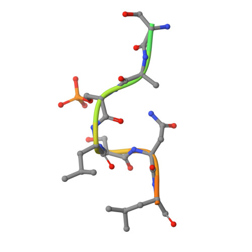
Crystal Structure of 14-3-3 zeta with Chibby peptide


Experimental Data Snapshot

  • Method: X-RAY DIFFRACTION
  • Resolution: 2.41 Å
  • R-Value Free: 0.256 
  • R-Value Work: 0.203 
  • R-Value Observed: 0.207 

wwPDB Validation   3D Report Full Report


This is version 1.3 of the entry. See complete history


Literature

Structural Analysis of the 14-3-3 zeta /Chibby Interaction Involved in Wnt/ beta-Catenin Signaling.

Killoran, R.C.Fan, J.Yang, D.Shilton, B.H.Choy, W.Y.

(2015) PLoS One 10: e0123934-e0123934

  • DOI: https://doi.org/10.1371/journal.pone.0123934
  • Primary Citation of Related Structures:  
    4WRQ

  • PubMed Abstract: 

    The partially disordered Chibby (Cby) is a conserved nuclear protein that antagonizes the Wnt/β-catenin signaling pathway. By competing with the Tcf/Lef family proteins for binding to β-catenin, Cby abrogates the β-catenin-mediated transcription of Wnt signaling genes. Additionally, upon phosphorylation on S20 by the kinase Akt, Cby forms a complex with 14-3-3 to facilitate the nuclear export of β-catenin, which represents another crucial mechanism for the regulation of Wnt signaling. To obtain a mechanistic understanding of the 14-3-3/Cby interaction, we have extensively characterized the complex using X-ray crystallography, nuclear magnetic resonance (NMR) spectroscopy, and isothermal titration calorimetry (ITC). The crystal structure of the human 14-3-3ζ/Cby protein-peptide complex reveals a canonical binding mode; however the residue at the +2 position from the phosphorylated serine is shown to be uniquely oriented relative to other solved structures of 14-3-3 complexes. Our ITC results illustrate that although the phosphorylation of S20 is essential for Cby to recognize 14-3-3, residues flanking the phosphorylation site also contribute to the binding affinity. However, as is commonly observed in other 14-3-3/phosphopeptide crystal structures, residues of Cby flanking the 14-3-3 binding motif lack observable electron density. To obtain a more detailed binding interface, we have completed the backbone NMR resonance assignment of 14-3-3ζ. NMR titration experiments reveal that residues outside of the 14-3-3 conserved binding cleft, namely a flexible loop consisting of residues 203-210, are also involved in binding Cby. By using a combined X-ray and NMR approach, we have dissected the molecular basis of the 14-3-3/Cby interaction.


  • Organizational Affiliation

    Department of Biochemistry, The University of Western Ontario, London, Ontario N6A 5C1, Canada.


Macromolecules
Find similar proteins by:  (by identity cutoff)  |  3D Structure
Entity ID: 1
MoleculeChains Sequence LengthOrganismDetailsImage
14-3-3 protein zeta/delta
A, B
246Homo sapiensMutation(s): 0 
Gene Names: YWHAZ
UniProt & NIH Common Fund Data Resources
Find proteins for P63104 (Homo sapiens)
Explore P63104 
Go to UniProtKB:  P63104
PHAROS:  P63104
GTEx:  ENSG00000164924 
Entity Groups  
Sequence Clusters30% Identity50% Identity70% Identity90% Identity95% Identity100% Identity
UniProt GroupP63104
Sequence Annotations
Expand
  • Reference Sequence

Find similar proteins by:  Sequence   |   3D Structure  

Entity ID: 2
MoleculeChains Sequence LengthOrganismDetailsImage
Chibby
C, D
18Homo sapiensMutation(s): 0 
UniProt & NIH Common Fund Data Resources
Find proteins for Q9Y3M2 (Homo sapiens)
Explore Q9Y3M2 
Go to UniProtKB:  Q9Y3M2
PHAROS:  Q9Y3M2
GTEx:  ENSG00000100211 
Entity Groups  
Sequence Clusters30% Identity50% Identity70% Identity90% Identity95% Identity100% Identity
UniProt GroupQ9Y3M2
Sequence Annotations
Expand
  • Reference Sequence
Small Molecules
Modified Residues  1 Unique
IDChains TypeFormula2D DiagramParent
SEP
Query on SEP
C, D
L-PEPTIDE LINKINGC3 H8 N O6 PSER
Experimental Data & Validation

Experimental Data

  • Method: X-RAY DIFFRACTION
  • Resolution: 2.41 Å
  • R-Value Free: 0.256 
  • R-Value Work: 0.203 
  • R-Value Observed: 0.207 
  • Space Group: P 21 21 21
Unit Cell:
Length ( Å )Angle ( ˚ )
a = 70.83α = 90
b = 71.96β = 90
c = 130.99γ = 90
Software Package:
Software NamePurpose
PHENIXrefinement
PDB_EXTRACTdata extraction

Structure Validation

View Full Validation Report


Entry History & Funding Information

Deposition Data


Funding OrganizationLocationGrant Number
Canadian Institutes of Health Research (CIHR)CanadaMOP 74679

Revision History  (Full details and data files)

  • Version 1.0: 2015-05-06
    Type: Initial release
  • Version 1.1: 2017-09-13
    Changes: Author supporting evidence, Derived calculations, Other, Source and taxonomy, Structure summary
  • Version 1.2: 2020-01-08
    Changes: Author supporting evidence
  • Version 1.3: 2023-09-27
    Changes: Data collection, Database references, Refinement description