Comparison of Saccharomyces cerevisiae F-BAR Domain Structures Reveals a Conserved Inositol Phosphate Binding Site.
Moravcevic, K., Alvarado, D., Schmitz, K.R., Kenniston, J.A., Mendrola, J.M., Ferguson, K.M., Lemmon, M.A.(2015) Structure 23: 352-363
- PubMed: 25620000
- DOI: https://doi.org/10.1016/j.str.2014.12.009
- Primary Citation of Related Structures:
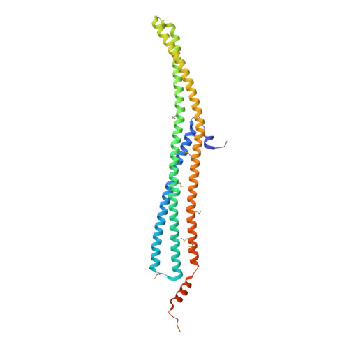
4WPC, 4WPE - PubMed Abstract:
F-BAR domains control membrane interactions in endocytosis, cytokinesis, and cell signaling. Although they are generally thought to bind curved membranes containing negatively charged phospholipids, numerous functional studies argue that differences in lipid-binding selectivities of F-BAR domains are functionally important. Here, we compare membrane-binding properties of the Saccharomyces cerevisiae F-BAR domains in vitro and in vivo. Whereas some F-BAR domains (such as Bzz1p and Hof1p F-BARs) bind equally well to all phospholipids, the F-BAR domain from the RhoGAP Rgd1p preferentially binds phosphoinositides. We determined X-ray crystal structures of F-BAR domains from Hof1p and Rgd1p, the latter bound to an inositol phosphate. The structures explain phospholipid-binding selectivity differences and reveal an F-BAR phosphoinositide binding site that is fully conserved in a mammalian RhoGAP called Gmip and is partly retained in certain other F-BAR domains. Our findings reveal previously unappreciated determinants of F-BAR domain lipid-binding specificity and provide a basis for its prediction from sequence.
Organizational Affiliation:
Department of Biochemistry and Biophysics, University of Pennsylvania Perelman School of Medicine, Philadelphia, PA 19014, USA; Graduate Group in Biochemistry and Molecular Biophysics, University of Pennsylvania Perelman School of Medicine, Philadelphia, PA 19014, USA.