4WO7

Crystal Structure of PrsA from Bacillus subtilis


Experimental Data Snapshot

  • Method: X-RAY DIFFRACTION
  • Resolution: 2.63 Å
  • R-Value Free: 0.251 
  • R-Value Work: 0.226 
  • R-Value Observed: 0.228 

wwPDB Validation   3D Report Full Report


This is version 2.0 of the entry. See complete history


Literature

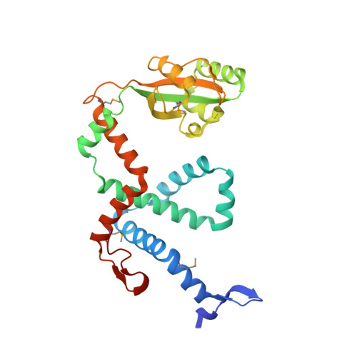
Dimeric Structure of the Bacterial Extracellular Foldase PrsA.

Jakob, R.P.Koch, J.R.Burmann, B.M.Schmidpeter, P.A.Hunkeler, M.Hiller, S.Schmid, F.X.Maier, T.

(2015) J Biol Chem 290: 3278-3292

  • DOI: https://doi.org/10.1074/jbc.M114.622910
  • Primary Citation of Related Structures:  
    4WO7

  • PubMed Abstract: 

    Secretion of proteins into the membrane-cell wall space is essential for cell wall biosynthesis and pathogenicity in Gram-positive bacteria. Folding and maturation of many secreted proteins depend on a single extracellular foldase, the PrsA protein. PrsA is a 30-kDa protein, lipid anchored to the outer leaflet of the cell membrane. The crystal structure of Bacillus subtilis PrsA reveals a central catalytic parvulin-type prolyl isomerase domain, which is inserted into a larger composite NC domain formed by the N- and C-terminal regions. This domain architecture resembles, despite a lack of sequence conservation, both trigger factor, a ribosome-binding bacterial chaperone, and SurA, a periplasmic chaperone in Gram-negative bacteria. Two main structural differences are observed in that the N-terminal arm of PrsA is substantially shortened relative to the trigger factor and SurA and in that PrsA is found to dimerize in a unique fashion via its NC domain. Dimerization leads to a large, bowl-shaped crevice, which might be involved in vivo in protecting substrate proteins from aggregation. NMR experiments reveal a direct, dynamic interaction of both the parvulin and the NC domain with secretion propeptides, which have been implicated in substrate targeting to PrsA.


  • Organizational Affiliation

    From the Biozentrum, Universität Basel, Klingelbergstrasse 50/70, 4056 Basel, Switzerland and roman.jakob@unibas.ch.


Macromolecules
Find similar proteins by:  (by identity cutoff)  |  3D Structure
Entity ID: 1
MoleculeChains Sequence LengthOrganismDetailsImage
Foldase protein PrsA
A, B
263Bacillus subtilis subsp. subtilis str. 168Mutation(s): 0 
Gene Names: prsABSU09950
EC: 5.2.1.8
UniProt
Find proteins for P24327 (Bacillus subtilis (strain 168))
Explore P24327 
Go to UniProtKB:  P24327
Entity Groups  
Sequence Clusters30% Identity50% Identity70% Identity90% Identity95% Identity100% Identity
UniProt GroupP24327
Sequence Annotations
Expand
  • Reference Sequence
Small Molecules
Modified Residues  1 Unique
IDChains TypeFormula2D DiagramParent
MSE
Query on MSE
A, B
L-PEPTIDE LINKINGC5 H11 N O2 SeMET
Experimental Data & Validation

Experimental Data

  • Method: X-RAY DIFFRACTION
  • Resolution: 2.63 Å
  • R-Value Free: 0.251 
  • R-Value Work: 0.226 
  • R-Value Observed: 0.228 
  • Space Group: C 2 2 21
Unit Cell:
Length ( Å )Angle ( ˚ )
a = 69.29α = 90
b = 87.68β = 90
c = 234.1γ = 90
Software Package:
Software NamePurpose
PHENIXrefinement
XSCALEdata scaling
XDSdata reduction
PHENIXphasing

Structure Validation

View Full Validation Report


Entry History & Funding Information

Deposition Data


Funding OrganizationLocationGrant Number
Swiss National Science FoundationSwitzerlandPP00P3_152989
Swiss National Science FoundationSwitzerlandCRSII3_147646

Revision History  (Full details and data files)

  • Version 1.0: 2014-12-24
    Type: Initial release
  • Version 1.1: 2014-12-31
    Changes: Database references
  • Version 1.2: 2015-02-18
    Changes: Database references
  • Version 2.0: 2017-09-13
    Changes: Advisory, Atomic model, Author supporting evidence