4WNK

Crystal Structure of Bovine G Protein Coupled-Receptor Kinase 5 in Complex with CCG215022

  • Classification: LIGASE
  • Organism(s): Bos taurus
  • Expression System: Trichoplusia ni
  • Mutation(s): No 

  • Deposited: 2014-10-13 Released: 2015-06-10 
  • Deposition Author(s): Homan, K.T., Tesmer, J.J.G.
  • Funding Organization(s): National Institutes of Health/National Heart, Lung, and Blood Institute (NIH/NHLBI), American Heart Association

Experimental Data Snapshot

  • Method: X-RAY DIFFRACTION
  • Resolution: 2.42 Å
  • R-Value Free: 0.243 
  • R-Value Work: 0.194 
  • R-Value Observed: 0.196 

wwPDB Validation   3D Report Full Report


Ligand Structure Quality Assessment 


This is version 1.6 of the entry. See complete history


Literature

Crystal Structure of G Protein-coupled Receptor Kinase 5 in Complex with a Rationally Designed Inhibitor.

Homan, K.T.Waldschmidt, H.V.Glukhova, A.Cannavo, A.Song, J.Cheung, J.Y.Koch, W.J.Larsen, S.D.Tesmer, J.J.

(2015) J Biol Chem 290: 20649-20659

  • DOI: https://doi.org/10.1074/jbc.M115.647370
  • Primary Citation of Related Structures:  
    4WNK

  • PubMed Abstract: 

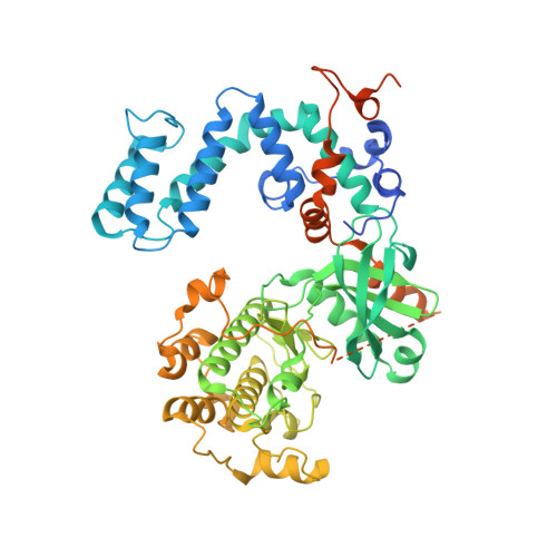
    G protein-coupled receptor kinases (GRKs) regulate cell signaling by initiating the desensitization of active G protein-coupled receptors. The two most widely expressed GRKs (GRK2 and GRK5) play a role in cardiovascular disease and thus represent important targets for the development of novel therapeutic drugs. In the course of a GRK2 structure-based drug design campaign, one inhibitor (CCG215022) exhibited nanomolar IC50 values against both GRK2 and GRK5 and good selectivity against other closely related kinases such as GRK1 and PKA. Treatment of murine cardiomyocytes with CCG215022 resulted in significantly increased contractility at 20-fold lower concentrations than paroxetine, an inhibitor with more modest selectivity for GRK2. A 2.4 Å crystal structure of the GRK5·CCG215022 complex was determined and revealed that the inhibitor binds in the active site similarly to its parent compound GSK180736A. As designed, its 2-pyridylmethyl amide side chain occupies the hydrophobic subsite of the active site where it forms three additional hydrogen bonds, including one with the catalytic lysine. The overall conformation of the GRK5 kinase domain is similar to that of a previously determined structure of GRK6 in what is proposed to be its active state, but the C-terminal region of the enzyme adopts a distinct conformation. The kinetic properties of site-directed mutants in this region are consistent with the hypothesis that this novel C-terminal structure is representative of the membrane-bound conformation of the enzyme.


  • Organizational Affiliation

    Life Sciences Institute and the Departments of Pharmacology and Biological Sciences, University of Michigan, Ann Arbor, Michigan 48109.


Macromolecules
Find similar proteins by:  (by identity cutoff)  |  3D Structure
Entity ID: 1
MoleculeChains Sequence LengthOrganismDetailsImage
G protein-coupled receptor kinase 5598Bos taurusMutation(s): 0 
Gene Names: GRK5GPRK5
EC: 2.7.11.16
UniProt
Find proteins for P43249 (Bos taurus)
Explore P43249 
Go to UniProtKB:  P43249
Entity Groups  
Sequence Clusters30% Identity50% Identity70% Identity90% Identity95% Identity100% Identity
UniProt GroupP43249
Sequence Annotations
Expand
  • Reference Sequence
Small Molecules
Ligands 2 Unique
IDChains Name / Formula / InChI Key2D Diagram3D Interactions
453
Query on 453

Download Ideal Coordinates CCD File 
B [auth A](4S)-4-{4-fluoro-3-[(pyridin-2-ylmethyl)carbamoyl]phenyl}-N-(1H-indazol-5-yl)-6-methyl-2-oxo-1,2,3,4-tetrahydropyrimidine-5-carboxamide
C26 H22 F N7 O3
BLMBNKDQXGINRE-QHCPKHFHSA-N
SO4
Query on SO4

Download Ideal Coordinates CCD File 
C [auth A]SULFATE ION
O4 S
QAOWNCQODCNURD-UHFFFAOYSA-L
Binding Affinity Annotations 
IDSourceBinding Affinity
453 BindingDB:  4WNK IC50: min: 280, max: 380 (nM) from 2 assay(s)
Binding MOAD:  4WNK IC50: 380 (nM) from 1 assay(s)
Experimental Data & Validation

Experimental Data

  • Method: X-RAY DIFFRACTION
  • Resolution: 2.42 Å
  • R-Value Free: 0.243 
  • R-Value Work: 0.194 
  • R-Value Observed: 0.196 
  • Space Group: P 21 21 21
Unit Cell:
Length ( Å )Angle ( ˚ )
a = 48.106α = 90
b = 70.104β = 90
c = 182.825γ = 90
Software Package:
Software NamePurpose
REFMACrefinement
HKL-2000data scaling
PHASERphasing

Structure Validation

View Full Validation Report


Ligand Structure Quality Assessment 


Entry History & Funding Information

Deposition Data


Funding OrganizationLocationGrant Number
National Institutes of Health/National Heart, Lung, and Blood Institute (NIH/NHLBI)United StatesHL071818
National Institutes of Health/National Heart, Lung, and Blood Institute (NIH/NHLBI)United StatesHL086865
American Heart AssociationUnited StatesN014938

Revision History  (Full details and data files)

  • Version 1.0: 2015-06-10
    Type: Initial release
  • Version 1.1: 2015-06-17
    Changes: Database references
  • Version 1.2: 2015-09-02
    Changes: Database references
  • Version 1.3: 2017-09-06
    Changes: Author supporting evidence, Database references, Derived calculations
  • Version 1.4: 2017-11-01
    Changes: Author supporting evidence, Refinement description
  • Version 1.5: 2019-12-04
    Changes: Author supporting evidence
  • Version 1.6: 2023-09-27
    Changes: Data collection, Database references, Refinement description