4WLR

Crystal Structure of mUCH37-hRPN13 CTD-hUb complex

  • Classification: PROTEIN BINDING
  • Organism(s): Mus musculus, Homo sapiens
  • Expression System: Escherichia coli
  • Mutation(s): No 

  • Deposited: 2014-10-07 Released: 2015-03-04 
  • Deposition Author(s): Hemmis, C.W., Hill, C.P., VanderLinden, R., Whitby, F.G.
  • Funding Organization(s): National Institutes of Health/National Institute of General Medical Sciences (NIH/NIGMS), National Institutes of Health/National Cancer Institute (NIH/NCI), National Institutes of Health/National Center for Research Resources (NIH/NCRR), Department of Energy (DOE, United States)

Experimental Data Snapshot

  • Method: X-RAY DIFFRACTION
  • Resolution: 2.00 Å
  • R-Value Free: 0.227 
  • R-Value Work: 0.181 
  • R-Value Observed: 0.183 

wwPDB Validation   3D Report Full Report


This is version 1.5 of the entry. See complete history


Literature

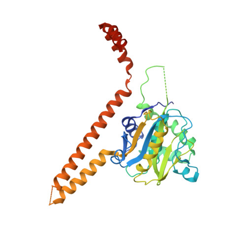
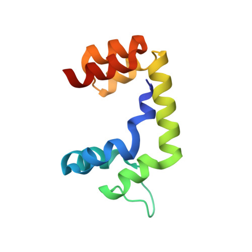
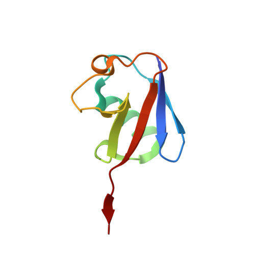
Structural Basis for the Activation and Inhibition of the UCH37 Deubiquitylase.

VanderLinden, R.T.Hemmis, C.W.Schmitt, B.Ndoja, A.Whitby, F.G.Robinson, H.Cohen, R.E.Yao, T.Hill, C.P.

(2015) Mol Cell 57: 901-911

  • DOI: https://doi.org/10.1016/j.molcel.2015.01.016
  • Primary Citation of Related Structures:  
    4WLP, 4WLQ, 4WLR

  • PubMed Abstract: 

    The UCH37 deubiquitylase functions in two large and very different complexes, the 26S proteasome and the INO80 chromatin remodeler. We have performed biochemical characterization and determined crystal structures of UCH37 in complexes with RPN13 and NFRKB, which mediate its recruitment to the proteasome and INO80, respectively. RPN13 and NFRKB make similar contacts to the UCH37 C-terminal domain but quite different contacts to the catalytic UCH domain. RPN13 can activate UCH37 by disrupting dimerization, although physiologically relevant activation likely results from stabilization of a surface competent for ubiquitin binding and modulation of the active-site crossover loop. In contrast, NFRKB inhibits UCH37 by blocking the ubiquitin-binding site and by disrupting the enzyme active site. These findings reveal remarkable commonality in mechanisms of recruitment, yet very different mechanisms of regulating enzyme activity, and provide a foundation for understanding the roles of UCH37 in the unrelated proteasome and INO80 complexes.


  • Organizational Affiliation

    Department of Biochemistry University of Utah School of Medicine, Salt Lake City, UT 84112-5650 USA.


Macromolecules
Find similar proteins by:  (by identity cutoff)  |  3D Structure
Entity ID: 1
MoleculeChains Sequence LengthOrganismDetailsImage
Ubiquitin carboxyl-terminal hydrolase isozyme L5328Mus musculusMutation(s): 0 
Gene Names: Uchl5Uch37
EC: 3.4.19.12
UniProt & NIH Common Fund Data Resources
Find proteins for Q9WUP7 (Mus musculus)
Explore Q9WUP7 
Go to UniProtKB:  Q9WUP7
IMPC:  MGI:1914848
Entity Groups  
Sequence Clusters30% Identity50% Identity70% Identity90% Identity95% Identity100% Identity
UniProt GroupQ9WUP7
Sequence Annotations
Expand
  • Reference Sequence
Find similar proteins by:  (by identity cutoff)  |  3D Structure
Entity ID: 2
MoleculeChains Sequence LengthOrganismDetailsImage
Proteasomal ubiquitin receptor ADRM1102Homo sapiensMutation(s): 0 
Gene Names: ADRM1GP110
UniProt & NIH Common Fund Data Resources
Find proteins for Q16186 (Homo sapiens)
Explore Q16186 
Go to UniProtKB:  Q16186
PHAROS:  Q16186
GTEx:  ENSG00000130706 
Entity Groups  
Sequence Clusters30% Identity50% Identity70% Identity90% Identity95% Identity100% Identity
UniProt GroupQ16186
Sequence Annotations
Expand
  • Reference Sequence
Find similar proteins by:  (by identity cutoff)  |  3D Structure
Entity ID: 3
MoleculeChains Sequence LengthOrganismDetailsImage
Polyubiquitin-B76Homo sapiensMutation(s): 0 
Gene Names: UBB
UniProt & NIH Common Fund Data Resources
Find proteins for P0CG47 (Homo sapiens)
Explore P0CG47 
Go to UniProtKB:  P0CG47
PHAROS:  P0CG47
GTEx:  ENSG00000170315 
Entity Groups  
Sequence Clusters30% Identity50% Identity70% Identity90% Identity95% Identity100% Identity
UniProt GroupP0CG47
Sequence Annotations
Expand
  • Reference Sequence
Experimental Data & Validation

Experimental Data

  • Method: X-RAY DIFFRACTION
  • Resolution: 2.00 Å
  • R-Value Free: 0.227 
  • R-Value Work: 0.181 
  • R-Value Observed: 0.183 
  • Space Group: P 21 21 21
Unit Cell:
Length ( Å )Angle ( ˚ )
a = 60.12α = 90
b = 99.61β = 90
c = 99.909γ = 90
Software Package:
Software NamePurpose
PHENIXrefinement
ADSCdata collection
XDSdata reduction
Aimlessdata scaling
PDB_EXTRACTdata extraction

Structure Validation

View Full Validation Report


Entry History & Funding Information

Deposition Data


Funding OrganizationLocationGrant Number
National Institutes of Health/National Institute of General Medical Sciences (NIH/NIGMS)United StatesRO1GM059135
National Institutes of Health/National Institute of General Medical Sciences (NIH/NIGMS)United StatesP50GM082545
National Institutes of Health/National Institute of General Medical Sciences (NIH/NIGMS)United StatesRO1GM098401
National Institutes of Health/National Institute of General Medical Sciences (NIH/NIGMS)United StatesRO1GM097452
National Institutes of Health/National Cancer Institute (NIH/NCI)United StatesP30CA042014
National Institutes of Health/National Institute of General Medical Sciences (NIH/NIGMS)United StatesP41GM103393
National Institutes of Health/National Center for Research Resources (NIH/NCRR)United StatesP41RR001209
Department of Energy (DOE, United States)United StatesDE-AC02-76SF00515

Revision History  (Full details and data files)

  • Version 1.0: 2015-03-04
    Type: Initial release
  • Version 1.1: 2015-03-11
    Changes: Database references, Structure summary
  • Version 1.2: 2015-03-25
    Changes: Database references
  • Version 1.3: 2017-09-20
    Changes: Author supporting evidence, Database references, Derived calculations, Other, Source and taxonomy
  • Version 1.4: 2019-12-04
    Changes: Author supporting evidence
  • Version 1.5: 2023-12-27
    Changes: Data collection, Database references, Refinement description