4WGK

Crystal structure of human neutral ceramidase with Zn-bound phosphate


Experimental Data Snapshot

  • Method: X-RAY DIFFRACTION
  • Resolution: 2.58 Å
  • R-Value Free: 0.223 
  • R-Value Work: 0.178 
  • R-Value Observed: 0.180 

wwPDB Validation   3D Report Full Report


Ligand Structure Quality Assessment 


This is version 2.1 of the entry. See complete history


Literature

Structural Basis for Ceramide Recognition and Hydrolysis by Human Neutral Ceramidase.

Airola, M.V.Allen, W.J.Pulkoski-Gross, M.J.Obeid, L.M.Rizzo, R.C.Hannun, Y.A.

(2015) Structure 23: 1482-1491

  • DOI: https://doi.org/10.1016/j.str.2015.06.013
  • Primary Citation of Related Structures:  
    4WGK

  • PubMed Abstract: 

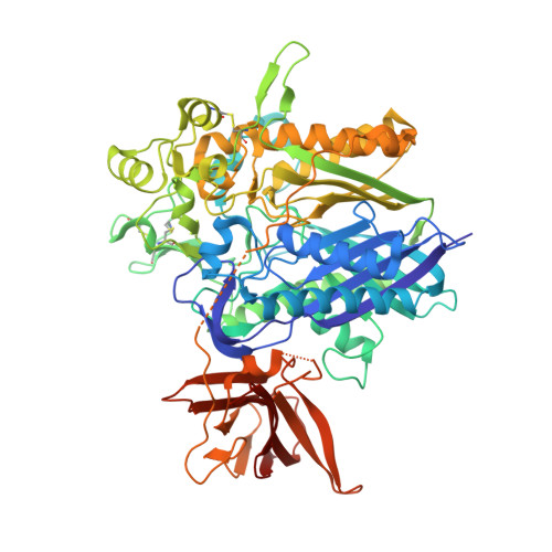
    Neutral ceramidase (nCDase) catalyzes conversion of the apoptosis-associated lipid ceramide to sphingosine, the precursor for the proliferative factor sphingosine-1-phosphate. As an enzyme regulating the balance of ceramide and sphingosine-1-phosphate, nCDase is emerging as a therapeutic target for cancer. Here, we present the 2.6-Å crystal structure of human nCDase in complex with phosphate that reveals a striking, 20-Å deep, hydrophobic active site pocket stabilized by a eukaryotic-specific subdomain not present in bacterial ceramidases. Utilizing flexible ligand docking, we predict a likely binding mode for ceramide that superimposes closely with the crystallographically observed transition state analog phosphate. Our results suggest that nCDase uses a new catalytic strategy for Zn(2+)-dependent amidases, and generates ceramide specificity by sterically excluding sphingolipids with bulky headgroups and specifically recognizing the small hydroxyl head group of ceramide. Together, these data provide a foundation to aid drug development and establish common themes for how proteins recognize the bioactive lipid ceramide.


  • Organizational Affiliation

    Department of Medicine, Stony Brook University, Stony Brook, NY 11794, USA; Department of Medicine, Stony Brook Cancer Center, Stony Brook, NY 11794, USA.


Macromolecules
Find similar proteins by:  (by identity cutoff)  |  3D Structure
Entity ID: 1
MoleculeChains Sequence LengthOrganismDetailsImage
Neutral ceramidase
A, B
688Homo sapiensMutation(s): 0 
Gene Names: ASAH2HNAC1
EC: 3.5.1.23
Membrane Entity: Yes 
UniProt & NIH Common Fund Data Resources
Find proteins for Q9NR71 (Homo sapiens)
Explore Q9NR71 
Go to UniProtKB:  Q9NR71
PHAROS:  Q9NR71
GTEx:  ENSG00000188611 
Entity Groups  
Sequence Clusters30% Identity50% Identity70% Identity90% Identity95% Identity100% Identity
UniProt GroupQ9NR71
Sequence Annotations
Expand
  • Reference Sequence
Oligosaccharides

Help

Entity ID: 2
MoleculeChains Length2D Diagram Glycosylation3D Interactions
2-acetamido-2-deoxy-beta-D-glucopyranose-(1-4)-2-acetamido-2-deoxy-beta-D-glucopyranose
C, E
2N-Glycosylation
Glycosylation Resources
GlyTouCan:  G42666HT
GlyCosmos:  G42666HT
GlyGen:  G42666HT
Entity ID: 3
MoleculeChains Length2D Diagram Glycosylation3D Interactions
beta-D-mannopyranose-(1-4)-2-acetamido-2-deoxy-beta-D-glucopyranose-(1-4)-2-acetamido-2-deoxy-beta-D-glucopyranose
D
3N-Glycosylation
Glycosylation Resources
GlyTouCan:  G15407YE
GlyCosmos:  G15407YE
GlyGen:  G15407YE
Small Molecules
Ligands 7 Unique
IDChains Name / Formula / InChI Key2D Diagram3D Interactions
NAG
Query on NAG

Download Ideal Coordinates CCD File 
H [auth A],
I [auth A],
O [auth B],
P [auth B],
Q [auth B]
2-acetamido-2-deoxy-beta-D-glucopyranose
C8 H15 N O6
OVRNDRQMDRJTHS-FMDGEEDCSA-N
CIT
Query on CIT

Download Ideal Coordinates CCD File 
K [auth A],
L [auth A],
T [auth B],
U [auth B]
CITRIC ACID
C6 H8 O7
KRKNYBCHXYNGOX-UHFFFAOYSA-N
PO4
Query on PO4

Download Ideal Coordinates CCD File 
J [auth A],
R [auth B]
PHOSPHATE ION
O4 P
NBIIXXVUZAFLBC-UHFFFAOYSA-K
ZN
Query on ZN

Download Ideal Coordinates CCD File 
F [auth A],
M [auth B]
ZINC ION
Zn
PTFCDOFLOPIGGS-UHFFFAOYSA-N
EDO
Query on EDO

Download Ideal Coordinates CCD File 
V [auth B],
W [auth B]
1,2-ETHANEDIOL
C2 H6 O2
LYCAIKOWRPUZTN-UHFFFAOYSA-N
CA
Query on CA

Download Ideal Coordinates CCD File 
G [auth A],
N [auth B]
CALCIUM ION
Ca
BHPQYMZQTOCNFJ-UHFFFAOYSA-N
CL
Query on CL

Download Ideal Coordinates CCD File 
S [auth B]CHLORIDE ION
Cl
VEXZGXHMUGYJMC-UHFFFAOYSA-M
Experimental Data & Validation

Experimental Data

  • Method: X-RAY DIFFRACTION
  • Resolution: 2.58 Å
  • R-Value Free: 0.223 
  • R-Value Work: 0.178 
  • R-Value Observed: 0.180 
  • Space Group: P 1 21 1
Unit Cell:
Length ( Å )Angle ( ˚ )
a = 73.48α = 90
b = 156.63β = 108.04
c = 80.33γ = 90
Software Package:
Software NamePurpose
PHENIXrefinement

Structure Validation

View Full Validation Report


Ligand Structure Quality Assessment 


Entry History & Funding Information

Deposition Data


Funding OrganizationLocationGrant Number
National Institutes of Health/National Cancer Institute (NIH/NCI)United StatesR01CA172517
National Institutes of Health/National Institute of General Medical Sciences (NIH/NIGMS)United StatesF32GM100679

Revision History  (Full details and data files)

  • Version 1.0: 2015-07-08
    Type: Initial release
  • Version 1.1: 2015-07-15
    Changes: Database references
  • Version 1.2: 2015-08-12
    Changes: Database references
  • Version 1.3: 2015-08-19
    Changes: Database references
  • Version 1.4: 2017-09-06
    Changes: Author supporting evidence, Database references, Derived calculations
  • Version 1.5: 2019-12-04
    Changes: Author supporting evidence
  • Version 2.0: 2020-07-29
    Type: Remediation
    Reason: Carbohydrate remediation
    Changes: Advisory, Atomic model, Data collection, Derived calculations, Structure summary
  • Version 2.1: 2023-09-27
    Changes: Data collection, Database references, Refinement description, Structure summary