4WEV

Crystal structure of human AKR1B10 complexed with NADP+ and sulindac


Experimental Data Snapshot

  • Method: X-RAY DIFFRACTION
  • Resolution: 1.45 Å
  • R-Value Free: 0.207 
  • R-Value Work: 0.188 
  • R-Value Observed: 0.189 

wwPDB Validation   3D Report Full Report


Ligand Structure Quality Assessment 


This is version 1.2 of the entry. See complete history


Literature

Structural analysis of sulindac as an inhibitor of aldose reductase and AKR1B10.

Cousido-Siah, A.Ruiz, F.X.Crespo, I.Porte, S.Mitschler, A.Pares, X.Podjarny, A.Farres, J.

(2015) Chem Biol Interact 234: 290-296

  • DOI: https://doi.org/10.1016/j.cbi.2014.12.018
  • Primary Citation of Related Structures:  
    4WEV

  • PubMed Abstract: 

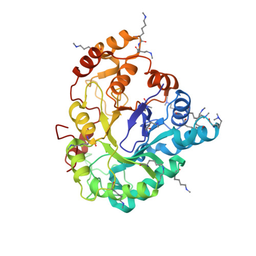
    Aldose reductase (AR, AKR1B1) and AKR1B10 are enzymes implicated in important pathologies (diabetes and cancer) and therefore they have been proposed as suitable targets for drug development. Sulindac is the metabolic precursor of the potent non-steroidal anti-inflammatory drug (NSAID) sulindac sulfide, which suppresses prostaglandin production by inhibition of cyclooxygenases (COX). In addition, sulindac has been found to be one of the NSAIDs with higher antitumoral activity, presumably through COX inhibition. However, sulindac anticancer activity could be partially mediated through COX-independent mechanisms, including the participation of AR and AKR1B10. Previously, it had been shown that sulindac and sulindac sulfone were good AR inhibitors and the structure of the ternary complex with NADP(+) and sulindac was described (PDB ID 3U2C). In this work, we determined the three-dimensional structure of AKR1B10 with sulindac and established structure-activity relationships (SAR) of sulindac and their derivatives with AR and AKR1B10. The difference in the IC50 values for sulindac between AR (0.36 μM) and AKR1B10 (2.7 μM) might be explained by the different positioning and stacking interaction given by Phe122/Phe123, and by the presence of two buried and ordered water molecules in AKR1B10 but not in AR. Moreover, SAR analysis shows that the substitution of the sulfinyl group is structurally allowed in sulindac derivatives. Hence, sulindac and its derivatives emerge as lead compounds for the design of more potent and selective AR and AKR1B10 inhibitors.


  • Organizational Affiliation

    Department of Integrative Structural Biology, Institut de Génétique et de Biologie Moléculaire et Cellulaire - Centre de Biologie Intégrative, CNRS, INSERM, UdS, 1 rue Laurent Fries, 67404 Illkirch Cedex, France.


Macromolecules
Find similar proteins by:  (by identity cutoff)  |  3D Structure
Entity ID: 1
MoleculeChains Sequence LengthOrganismDetailsImage
Aldo-keto reductase family 1 member B10A [auth X]316Homo sapiensMutation(s): 2 
Gene Names: AKR1B10AKR1B11
EC: 1.1.1
UniProt & NIH Common Fund Data Resources
Find proteins for O60218 (Homo sapiens)
Explore O60218 
Go to UniProtKB:  O60218
PHAROS:  O60218
GTEx:  ENSG00000198074 
Entity Groups  
Sequence Clusters30% Identity50% Identity70% Identity90% Identity95% Identity100% Identity
UniProt GroupO60218
Sequence Annotations
Expand
  • Reference Sequence
Small Molecules
Ligands 2 Unique
IDChains Name / Formula / InChI Key2D Diagram3D Interactions
NAP
Query on NAP

Download Ideal Coordinates CCD File 
B [auth X]NADP NICOTINAMIDE-ADENINE-DINUCLEOTIDE PHOSPHATE
C21 H28 N7 O17 P3
XJLXINKUBYWONI-NNYOXOHSSA-N
SUZ
Query on SUZ

Download Ideal Coordinates CCD File 
C [auth X][(1Z)-5-fluoro-2-methyl-1-{4-[methylsulfinyl]benzylidene}-1H-inden-3-yl]acetic acid
C20 H17 F O3 S
MLKXDPUZXIRXEP-LQVWSKNFSA-N
Modified Residues  2 Unique
IDChains TypeFormula2D DiagramParent
MLY
Query on MLY
A [auth X]L-PEPTIDE LINKINGC8 H18 N2 O2LYS
MLZ
Query on MLZ
A [auth X]L-PEPTIDE LINKINGC7 H16 N2 O2LYS
Binding Affinity Annotations 
IDSourceBinding Affinity
SUZ BindingDB:  4WEV IC50: 2690 (nM) from 1 assay(s)
Experimental Data & Validation

Experimental Data

  • Method: X-RAY DIFFRACTION
  • Resolution: 1.45 Å
  • R-Value Free: 0.207 
  • R-Value Work: 0.188 
  • R-Value Observed: 0.189 
  • Space Group: P 31
Unit Cell:
Length ( Å )Angle ( ˚ )
a = 78.848α = 90
b = 78.848β = 90
c = 50.127γ = 120
Software Package:
Software NamePurpose
HKL-2000data reduction
HKL-2000data scaling
Cootmodel building
PHASERphasing
PHENIXrefinement

Structure Validation

View Full Validation Report


Ligand Structure Quality Assessment 


Entry History 

Deposition Data

Revision History  (Full details and data files)

  • Version 1.0: 2015-01-14
    Type: Initial release
  • Version 1.1: 2015-05-13
    Changes: Database references
  • Version 1.2: 2024-01-10
    Changes: Data collection, Database references, Refinement description