In-Cell Enzymology To Probe His-Heme Ligation in Heme Oxygenase Catalysis
Sigala, P.A., Morante, K., Tsumoto, K., Caaveiro, J.M., Goldberg, D.E.(2016) Biochemistry 55: 4836-4849
- PubMed: 27490825
- DOI: https://doi.org/10.1021/acs.biochem.6b00562
- Primary Citation of Related Structures:
4WD4, 5BTQ - PubMed Abstract:
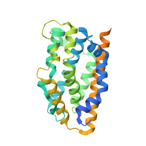
Heme oxygenase (HO) is a ubiquitous enzyme with key roles in inflammation, cell signaling, heme disposal, and iron acquisition. HO catalyzes the oxidative conversion of heme to biliverdin (BV) using a conserved histidine to coordinate the iron atom of bound heme. This His-heme interaction has been regarded as being essential for enzyme activity, because His-to-Ala mutants fail to convert heme to biliverdin in vitro. We probed a panel of proximal His mutants of cyanobacterial, human, and plant HO enzymes using a live-cell activity assay based on heterologous co-expression in Escherichia coli of each HO mutant and a fluorescent biliverdin biosensor. In contrast to in vitro studies with purified proteins, we observed that multiple HO mutants retained significant activity within the intracellular environment of bacteria. X-ray crystallographic structures of human HO1 H25R with bound heme and additional functional studies suggest that HO mutant activity inside these cells does not involve heme ligation by a proximal amino acid. Our study reveals unexpected plasticity in the active site binding interactions with heme that can support HO activity within cells, suggests important contributions by the surrounding active site environment to HO catalysis, and can guide efforts to understand the evolution and divergence of HO function.
Organizational Affiliation:
Departments of Medicine and Molecular Microbiology, Washington University School of Medicine , St. Louis, Missouri 63110, United States.