4V24

Sphingosine kinase 1 in complex with PF-543


Experimental Data Snapshot

  • Method: X-RAY DIFFRACTION
  • Resolution: 1.80 Å
  • R-Value Free: 0.210 
  • R-Value Work: 0.182 
  • R-Value Observed: 0.183 

wwPDB Validation   3D Report Full Report


Ligand Structure Quality Assessment 


This is version 1.3 of the entry. See complete history


Literature

Crystal Structure of Sphingosine Kinase 1 with Pf-543.

Wang, J.Knapp, S.Pyne, N.J.Pyne, S.Elkins, J.M.

(2014) ACS Med Chem Lett 5: 1329

  • DOI: https://doi.org/10.1021/ml5004074
  • Primary Citation of Related Structures:  
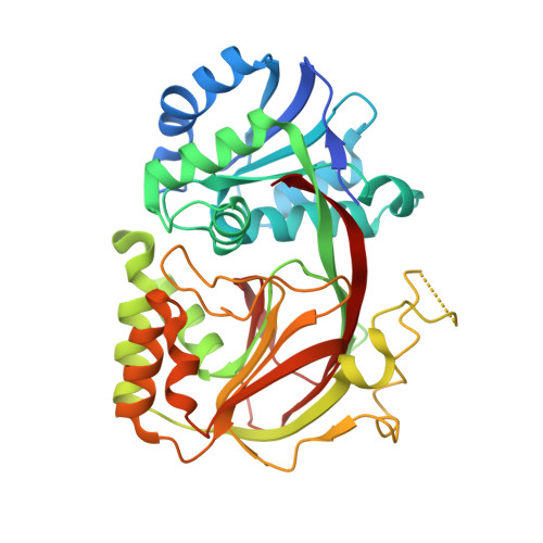
    4V24

  • PubMed Abstract: 

    The most potent inhibitor of Sphingosine Kinase 1 (SPHK1) so far identified is PF-543. The crystal structure of SPHK1 in complex with inhibitor PF-543 to 1.8 Å resolution reveals the inhibitor bound in a bent conformation analogous to that expected of a bound sphingosine substrate but with a rotated head group. The structural data presented will aid in the design of SPHK1 and SPHK2 inhibitors with improved properties.


  • Organizational Affiliation

    Structural Genomics Consortium, University of Oxford , Old Road Campus Research Building, Old Road Campus, Roosevelt Drive, Oxford OX3 7DQ, U.K.


Macromolecules
Find similar proteins by:  (by identity cutoff)  |  3D Structure
Entity ID: 1
MoleculeChains Sequence LengthOrganismDetailsImage
SPHINGOSINE KINASE 1
A, B
371Homo sapiensMutation(s): 0 
EC: 2.7.1.91
UniProt & NIH Common Fund Data Resources
Find proteins for Q9NYA1 (Homo sapiens)
Explore Q9NYA1 
Go to UniProtKB:  Q9NYA1
PHAROS:  Q9NYA1
GTEx:  ENSG00000176170 
Entity Groups  
Sequence Clusters30% Identity50% Identity70% Identity90% Identity95% Identity100% Identity
UniProt GroupQ9NYA1
Sequence Annotations
Expand
  • Reference Sequence
Small Molecules
Ligands 2 Unique
IDChains Name / Formula / InChI Key2D Diagram3D Interactions
GYR
Query on GYR

Download Ideal Coordinates CCD File 
C [auth A],
E [auth B]
{(2R)-1-[4-({3-METHYL-5-[(PHENYLSULFONYL)METHYL]PHENOXY}METHYL)BENZYL]PYRROLIDIN-2-YL}METHANOL
C27 H31 N O4 S
NPUXORBZRBIOMQ-RUZDIDTESA-N
ACT
Query on ACT

Download Ideal Coordinates CCD File 
D [auth A]ACETATE ION
C2 H3 O2
QTBSBXVTEAMEQO-UHFFFAOYSA-M
Binding Affinity Annotations 
IDSourceBinding Affinity
GYR Binding MOAD:  4V24 Ki: 4.3 (nM) from 1 assay(s)
BindingDB:  4V24 Ki: min: 3.6, max: 4.3 (nM) from 3 assay(s)
Kd: 5 (nM) from 1 assay(s)
IC50: min: 0.8, max: 387 (nM) from 8 assay(s)
Experimental Data & Validation

Experimental Data

  • Method: X-RAY DIFFRACTION
  • Resolution: 1.80 Å
  • R-Value Free: 0.210 
  • R-Value Work: 0.182 
  • R-Value Observed: 0.183 
  • Space Group: P 1 21 1
Unit Cell:
Length ( Å )Angle ( ˚ )
a = 86.57α = 90
b = 60.549β = 107.17
c = 89.592γ = 90
Software Package:
Software NamePurpose
REFMACrefinement
XDSdata reduction
Aimlessdata scaling
PHASERphasing

Structure Validation

View Full Validation Report


Ligand Structure Quality Assessment 


Entry History 

Deposition Data

Revision History  (Full details and data files)

  • Version 1.0: 2014-10-15
    Type: Initial release
  • Version 1.1: 2014-12-31
    Changes: Database references
  • Version 1.2: 2018-01-24
    Changes: Structure summary
  • Version 1.3: 2024-01-10
    Changes: Data collection, Database references, Derived calculations, Other, Refinement description