4UU1

CRYSTAL STRUCTURE OF (SR) CALCIUM-ATPASE E2(TG) IN THE PRESENCE OF DOPC


Experimental Data Snapshot

  • Method: X-RAY DIFFRACTION
  • Resolution: 2.80 Å
  • R-Value Free: 0.293 
  • R-Value Work: 0.221 
  • R-Value Observed: 0.225 

wwPDB Validation   3D Report Full Report


Ligand Structure Quality Assessment 


This is version 1.2 of the entry. See complete history


Literature

Comparing Crystal Structures of Ca(2+) -ATPase in the Presence of Different Lipids.

Drachmann, N.D.Olesen, C.Moller, J.V.Guo, Z.Nissen, P.Bublitz, M.

(2014) FEBS J 281: 4249

  • DOI: https://doi.org/10.1111/febs.12957
  • Primary Citation of Related Structures:  
    4UU0, 4UU1

  • PubMed Abstract: 

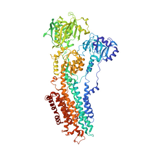
    The activity of the sarco/endoplasmic reticulum Ca(2+) -ATPase (SERCA) depends strongly on the lipid composition of the surrounding membrane. Yet, structural information on SERCA-lipid interaction is still relatively scarce, and the influence of different lipids on the enzyme is not well understood. We have analyzed SERCA crystal structures in the presence of four different phosphatidylcholine lipids of different lengths and double-bond compositions, and we find three different binding sites for lipid head groups, which are apparently independent of the acyl moiety of the lipids used. By comparison with other available SERCA structures with bound lipids, we find a total of five recurring sites, two of which are specific to certain conformational states of the enzyme, two others are state-independent, and one is a crucial site for crystal formation. Three of the binding sites overlap with or are in close vicinity to known binding sites for various SERCA-specific inhibitors and regulators, e.g. thapsigargin, sarcolipin/phospholamban and cyclopiazonic acid. Whereas the transient sites are amenable to a transient, regulatory influence of lipid molecules, the state-independent sites probably provide a flexible anchoring of the protein in the fluid bilayer.


  • Organizational Affiliation

    Centre for Membrane Pumps in Cells and Disease - PUMPkin, Danish National Research Foundation, Aarhus, Denmark; Department of Molecular Biology and Genetics, Aarhus University, Denmark.


Macromolecules
Find similar proteins by:  (by identity cutoff)  |  3D Structure
Entity ID: 1
MoleculeChains Sequence LengthOrganismDetailsImage
SARCOPLASMIC ENDOPLASMIC RETICULUM CALCIUM ATPASE995Oryctolagus cuniculusMutation(s): 1 
EC: 3.6.3.8
Membrane Entity: Yes 
UniProt
Find proteins for P04191 (Oryctolagus cuniculus)
Explore P04191 
Go to UniProtKB:  P04191
Entity Groups  
Sequence Clusters30% Identity50% Identity70% Identity90% Identity95% Identity100% Identity
UniProt GroupP04191
Sequence Annotations
Expand
  • Reference Sequence
Small Molecules
Ligands 6 Unique
IDChains Name / Formula / InChI Key2D Diagram3D Interactions
PCW
Query on PCW

Download Ideal Coordinates CCD File 
C [auth A],
D [auth A],
E [auth A]
1,2-DIOLEOYL-SN-GLYCERO-3-PHOSPHOCHOLINE
C44 H85 N O8 P
SNKAWJBJQDLSFF-NVKMUCNASA-O
TG1
Query on TG1

Download Ideal Coordinates CCD File 
B [auth A]OCTANOIC ACID [3S-[3ALPHA, 3ABETA, 4ALPHA, 6BETA, 6ABETA, 7BETA, 8ALPHA(Z), 9BALPHA]]-6-(ACETYLOXY)-2,3,-3A,4,5,6,6A,7,8,9B-DECAHYDRO-3,3A-DIHYDROXY-3,6,9-TRIMETHYL-8-[(2-METHYL-1-OXO-2-BUTENYL)OX Y]-2-OXO-4-(1-OXOBUTOXY)-AZULENO[4,5-B]FURAN-7-YL ESTER
C34 H50 O12
IXFPJGBNCFXKPI-FSIHEZPISA-N
ACP
Query on ACP

Download Ideal Coordinates CCD File 
H [auth A]PHOSPHOMETHYLPHOSPHONIC ACID ADENYLATE ESTER
C11 H18 N5 O12 P3
UFZTZBNSLXELAL-IOSLPCCCSA-N
GOL
Query on GOL

Download Ideal Coordinates CCD File 
F [auth A],
G [auth A]
GLYCEROL
C3 H8 O3
PEDCQBHIVMGVHV-UHFFFAOYSA-N
K
Query on K

Download Ideal Coordinates CCD File 
I [auth A]POTASSIUM ION
K
NPYPAHLBTDXSSS-UHFFFAOYSA-N
MG
Query on MG

Download Ideal Coordinates CCD File 
J [auth A]MAGNESIUM ION
Mg
JLVVSXFLKOJNIY-UHFFFAOYSA-N
Binding Affinity Annotations 
IDSourceBinding Affinity
TG1 BindingDB:  4UU1 Ki: 0.1 (nM) from 1 assay(s)
Kd: 0.2 (nM) from 1 assay(s)
IC50: min: 0.2, max: 3.9 (nM) from 2 assay(s)
Experimental Data & Validation

Experimental Data

  • Method: X-RAY DIFFRACTION
  • Resolution: 2.80 Å
  • R-Value Free: 0.293 
  • R-Value Work: 0.221 
  • R-Value Observed: 0.225 
  • Space Group: P 41 21 2
Unit Cell:
Length ( Å )Angle ( ˚ )
a = 71.703α = 90
b = 71.703β = 90
c = 591.318γ = 90
Software Package:
Software NamePurpose
PHENIXrefinement
XDSdata reduction
XSCALEdata scaling
PHASERphasing

Structure Validation

View Full Validation Report


Ligand Structure Quality Assessment 


Entry History 

Deposition Data

Revision History  (Full details and data files)

  • Version 1.0: 2014-10-01
    Type: Initial release
  • Version 1.1: 2019-10-23
    Changes: Data collection, Database references, Derived calculations, Other
  • Version 1.2: 2024-01-10
    Changes: Data collection, Database references, Derived calculations, Refinement description, Structure summary