4UTQ

A structural model of the active ribosome-bound membrane protein insertase YidC


Experimental Data Snapshot

  • Method: ELECTRON MICROSCOPY
  • Resolution: 8.00 Å
  • Aggregation State: PARTICLE 
  • Reconstruction Method: SINGLE PARTICLE 

wwPDB Validation   3D Report Full Report


This is version 1.2 of the entry. See complete history


Literature

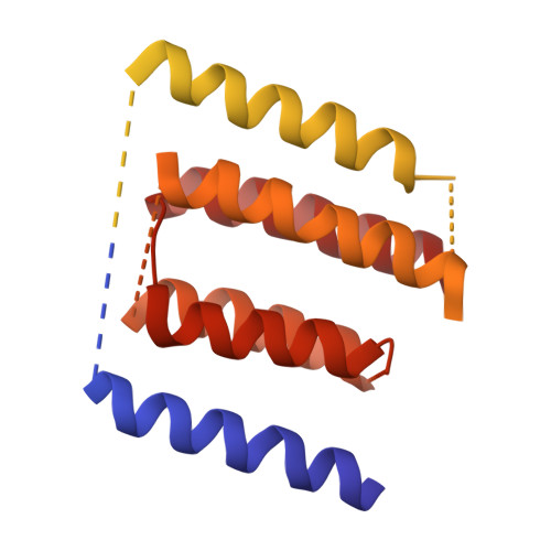
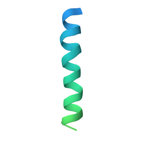
A Structural Model of the Active Ribosome-Bound Membrane Protein Insertase Yidc.

Wickles, S.Singharoy, A.Andreani, J.Seemayer, S.Bischoff, L.Berninghausen, O.Soeding, J.Schulten, K.Van Der Sluis, E.O.Beckmann, R.

(2014) Elife 3: 3035

  • DOI: https://doi.org/10.7554/eLife.03035
  • Primary Citation of Related Structures:  
    4UTQ

  • PubMed Abstract: 

    The integration of most membrane proteins into the cytoplasmic membrane of bacteria occurs co-translationally. The universally conserved YidC protein mediates this process either individually as a membrane protein insertase, or in concert with the SecY complex. Here, we present a structural model of YidC based on evolutionary co-variation analysis, lipid-versus-protein-exposure and molecular dynamics simulations. The model suggests a distinctive arrangement of the conserved five transmembrane domains and a helical hairpin between transmembrane segment 2 (TM2) and TM3 on the cytoplasmic membrane surface. The model was used for docking into a cryo-electron microscopy reconstruction of a translating YidC-ribosome complex carrying the YidC substrate FOc. This structure reveals how a single copy of YidC interacts with the ribosome at the ribosomal tunnel exit and identifies a site for membrane protein insertion at the YidC protein-lipid interface. Together, these data suggest a mechanism for the co-translational mode of YidC-mediated membrane protein insertion.


  • Organizational Affiliation

    Gene Center Munich, Department of Biochemistry, Ludwig-Maximilians-Universität München, Munich, Germany Center for Integrated Protein Science Munich, Department of Biochemistry, Ludwig-Maximilians-Universität München, Munich, Germany.


Macromolecules
Find similar proteins by:  (by identity cutoff)  |  3D Structure
Entity ID: 1
MoleculeChains Sequence LengthOrganismDetailsImage
MEMBRANE PROTEIN INSERTASE YIDC548Escherichia coliMutation(s): 0 
Membrane Entity: Yes 
UniProt
Find proteins for P25714 (Escherichia coli (strain K12))
Explore P25714 
Go to UniProtKB:  P25714
Entity Groups  
Sequence Clusters30% Identity50% Identity70% Identity90% Identity95% Identity100% Identity
UniProt GroupP25714
Sequence Annotations
Expand
  • Reference Sequence
Find similar proteins by:  (by identity cutoff)  |  3D Structure
Entity ID: 2
MoleculeChains Sequence LengthOrganismDetailsImage
ATP SYNTHASE SUBUNIT CB [auth Z]79Escherichia coliMutation(s): 0 
Membrane Entity: Yes 
UniProt
Find proteins for P68699 (Escherichia coli (strain K12))
Explore P68699 
Go to UniProtKB:  P68699
Entity Groups  
Sequence Clusters30% Identity50% Identity70% Identity90% Identity95% Identity100% Identity
UniProt GroupP68699
Sequence Annotations
Expand
  • Reference Sequence
Experimental Data & Validation

Experimental Data

  • Method: ELECTRON MICROSCOPY
  • Resolution: 8.00 Å
  • Aggregation State: PARTICLE 
  • Reconstruction Method: SINGLE PARTICLE 
EM Software:
TaskSoftware PackageVersion
RECONSTRUCTIONSPIDER

Structure Validation

View Full Validation Report


Entry History 

Revision History  (Full details and data files)

  • Version 1.0: 2014-07-30
    Type: Initial release
  • Version 1.1: 2014-08-20
    Changes: Database references
  • Version 1.2: 2018-10-03
    Changes: Data collection