Structure of the Bacterial Cell Division Determinant Gpsb and its Interaction with Penicillin Binding Proteins.
Rismondo, J., Cleverley, R.M., Lane, H.V., Grosshennig, S., Steglich, A., Moller, L., Mannala, G.K., Hain, T., Lewis, R.J., Halbedel, S.(2016) Mol Microbiol 99: 978
- PubMed: 26575090
- DOI: https://doi.org/10.1111/mmi.13279
- Primary Citation of Related Structures:
4UG1, 4UG3, 5AN5 - PubMed Abstract:
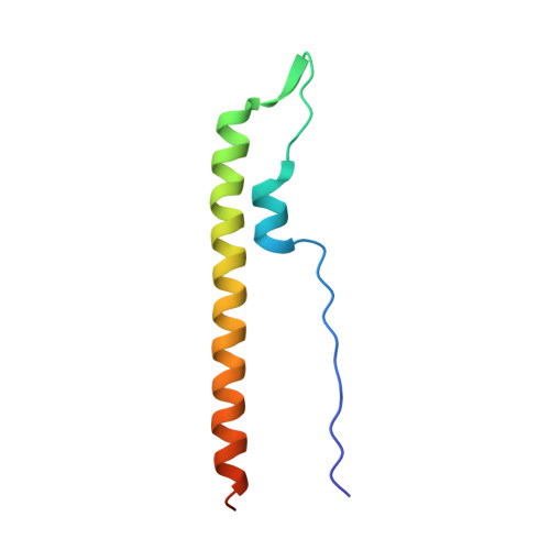
Each bacterium has to co-ordinate its growth with division to ensure genetic stability of the population. Consequently, cell division and growth are tightly regulated phenomena, albeit different bacteria utilise one of several alternative regulatory mechanisms to maintain control. Here we consider GpsB, which is linked to cell growth and division in Gram-positive bacteria. ΔgpsB mutants of the human pathogen Listeria monocytogenes show severe lysis, division and growth defects due to distortions of cell wall biosynthesis. Consistent with this premise, GpsB interacts both in vitro and in vivo with the major bi-functional penicillin-binding protein. We solved the crystal structure of GpsB and the interaction interfaces in both proteins are identified and validated. The inactivation of gpsB results in strongly attenuated virulence in animal experiments, comparable in degree to classical listerial virulence factor mutants. Therefore, GpsB is essential for in vitro and in vivo growth of a highly virulent food-borne pathogen, suggesting that GpsB could be a target for the future design of novel antibacterials.
Organizational Affiliation:
FG11 Division of Enteropathogenic Bacteria and Legionella, Robert Koch Institute, Wernigerode, Germany.