


Crystal Structure of the Human Carboxypeptidase A1 in Complex with Phosphinic Inhibitors
Gallego, P., Covaleda, G., Devel, L., Dive, V., Aviles, F.X., Reverter, D.To be published.
Experimental Data Snapshot
Entity ID: 1 | |||||
|---|---|---|---|---|---|
| Molecule | Chains | Sequence Length | Organism | Details | Image |
| HUMAN CARBOXYPEPTIDASE A1 | 309 | Homo sapiens | Mutation(s): 0 EC: 3.4.17.1 | 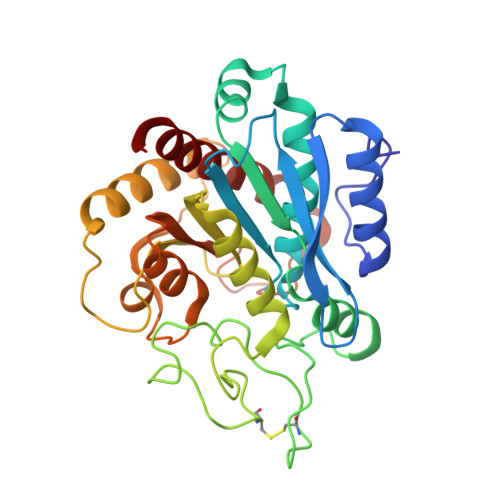 | |
UniProt & NIH Common Fund Data Resources | |||||
Find proteins for P15085 (Homo sapiens) Explore P15085 Go to UniProtKB: P15085 | |||||
PHAROS: P15085 GTEx: ENSG00000091704 | |||||
Entity Groups | |||||
| Sequence Clusters | 30% Identity50% Identity70% Identity90% Identity95% Identity100% Identity | ||||
| UniProt Group | P15085 | ||||
Sequence AnnotationsExpand | |||||
| |||||
| Ligands 2 Unique | |||||
|---|---|---|---|---|---|
| ID | Chains | Name / Formula / InChI Key | 2D Diagram | 3D Interactions | |
| LFF Query on LFF | D [auth A], F [auth B] | (2S)-3-[(R)-{(1R)-1-[(N-acetyl-L-leucyl)amino]-2-phenylethyl}(hydroxy)phosphoryl]-2-benzylpropanoic acid C26 H35 N2 O6 P GRMCSCYPDCPNRA-TZRRMPRUSA-N |  | ||
| ZN Query on ZN | C [auth A], E [auth B] | ZINC ION Zn PTFCDOFLOPIGGS-UHFFFAOYSA-N |  | ||
| Length ( Å ) | Angle ( ˚ ) |
|---|---|
| a = 45.989 | α = 90 |
| b = 84.143 | β = 90 |
| c = 158.005 | γ = 90 |
| Software Name | Purpose |
|---|---|
| REFMAC | refinement |
| XPS | data reduction |
| CCP4I | data scaling |
| MOLREP | phasing |
RCSB PDB (citation) is hosted by
RCSB PDB is a member of the

