


Crystal Structure of Human Glycogenin-2 Catalytic Domain
Fairhead, M., Strain-Damerell, C., Krojer, T., Froese, D.S., Kopec, J., Nowak, R., Burgess-Brown, N., von Delft, F., Arrowsmith, C., Edwards, A., Bountra, C., Yue, W.W.To be published.
Experimental Data Snapshot
wwPDB Validation 3D Report Full Report

Entity ID: 1 | |||||
|---|---|---|---|---|---|
| Molecule | Chains | Sequence Length | Organism | Details | Image |
| GLYCOGENIN-2 | 288 | Homo sapiens | Mutation(s): 2 EC: 2.4.1.186 | 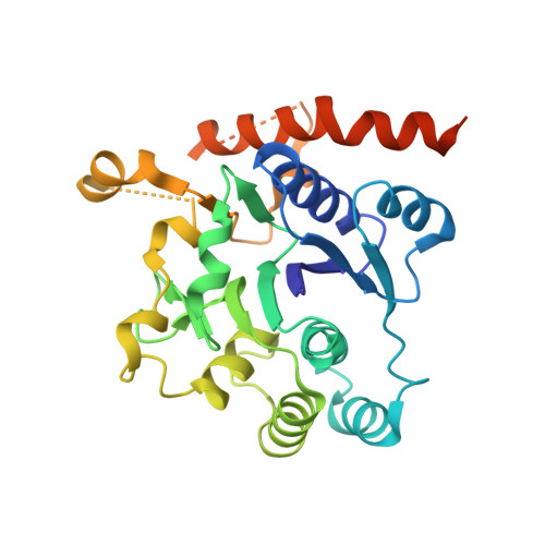 | |
UniProt & NIH Common Fund Data Resources | |||||
Find proteins for O15488 (Homo sapiens) Explore O15488 Go to UniProtKB: O15488 | |||||
PHAROS: O15488 GTEx: ENSG00000056998 | |||||
Entity Groups | |||||
| Sequence Clusters | 30% Identity50% Identity70% Identity90% Identity95% Identity100% Identity | ||||
| UniProt Group | O15488 | ||||
Sequence AnnotationsExpand | |||||
| |||||
| Ligands 1 Unique | |||||
|---|---|---|---|---|---|
| ID | Chains | Name / Formula / InChI Key | 2D Diagram | 3D Interactions | |
| MG Query on MG | C [auth A], D [auth B] | MAGNESIUM ION Mg JLVVSXFLKOJNIY-UHFFFAOYSA-N |  | ||
| Length ( Å ) | Angle ( ˚ ) |
|---|---|
| a = 127.113 | α = 90 |
| b = 80.541 | β = 118.74 |
| c = 73.683 | γ = 90 |
| Software Name | Purpose |
|---|---|
| PHENIX | refinement |
| XDS | data reduction |
| SCALEPACK | data scaling |
| PHASER | phasing |
RCSB PDB (citation) is hosted by
RCSB PDB is a member of the

