4UC3

Translocator protein 18 kDa (TSPO) from Rhodobacter sphaeroides wild type


Experimental Data Snapshot

  • Method: X-RAY DIFFRACTION
  • Resolution: 2.50 Å
  • R-Value Free: 0.278 
  • R-Value Work: 0.218 
  • R-Value Observed: 0.220 

wwPDB Validation   3D Report Full Report


Ligand Structure Quality Assessment 


This is version 1.6 of the entry. See complete history


Literature

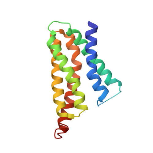
Crystal structures of translocator protein (TSPO) and mutant mimic of a human polymorphism.

Li, F.Liu, J.Zheng, Y.Garavito, R.M.Ferguson-Miller, S.

(2015) Science 347: 555-558

  • DOI: https://doi.org/10.1126/science.1260590
  • Primary Citation of Related Structures:  
    4UC1, 4UC2, 4UC3

  • PubMed Abstract: 

    The 18-kilodalton translocator protein (TSPO), proposed to be a key player in cholesterol transport into mitochondria, is highly expressed in steroidogenic tissues, metastatic cancer, and inflammatory and neurological diseases such as Alzheimer's and Parkinson's. TSPO ligands, including benzodiazepine drugs, are implicated in regulating apoptosis and are extensively used in diagnostic imaging. We report crystal structures (at 1.8, 2.4, and 2.5 angstrom resolution) of TSPO from Rhodobacter sphaeroides and a mutant that mimics the human Ala(147)→Thr(147) polymorphism associated with psychiatric disorders and reduced pregnenolone production. Crystals obtained in the lipidic cubic phase reveal the binding site of an endogenous porphyrin ligand and conformational effects of the mutation. The three crystal structures show the same tightly interacting dimer and provide insights into the controversial physiological role of TSPO and how the mutation affects cholesterol binding.


  • Organizational Affiliation

    Department of Biochemistry and Molecular Biology, Michigan State University, East Lansing, MI 48824, USA.


Macromolecules
Find similar proteins by:  (by identity cutoff)  |  3D Structure
Entity ID: 1
MoleculeChains Sequence LengthOrganismDetailsImage
TRANSLOCATOR PROTEIN TSPO
A, B
155Cereibacter sphaeroidesMutation(s): 0 
Membrane Entity: Yes 
UniProt
Find proteins for Q9RFC8 (Cereibacter sphaeroides)
Explore Q9RFC8 
Go to UniProtKB:  Q9RFC8
Entity Groups  
Sequence Clusters30% Identity50% Identity70% Identity90% Identity95% Identity100% Identity
UniProt GroupQ9RFC8
Sequence Annotations
Expand
  • Reference Sequence
Experimental Data & Validation

Experimental Data

  • Method: X-RAY DIFFRACTION
  • Resolution: 2.50 Å
  • R-Value Free: 0.278 
  • R-Value Work: 0.218 
  • R-Value Observed: 0.220 
  • Space Group: P 1 21 1
Unit Cell:
Length ( Å )Angle ( ˚ )
a = 49.1α = 90
b = 61.706β = 112.53
c = 52.676γ = 90
Software Package:
Software NamePurpose
Blu-Icedata collection
XDSdata scaling
Aimlessdata scaling
PHENIXrefinement

Structure Validation

View Full Validation Report


Ligand Structure Quality Assessment 


Entry History & Funding Information

Deposition Data


Funding OrganizationLocationGrant Number
National Institutes of Health/National Institute of General Medical Sciences (NIH/NIGMS)United StatesNIH GM26916

Revision History  (Full details and data files)

  • Version 1.0: 2015-02-04
    Type: Initial release
  • Version 1.1: 2015-02-11
    Changes: Database references
  • Version 1.2: 2015-06-03
    Changes: Structure summary
  • Version 1.3: 2017-09-13
    Changes: Author supporting evidence, Database references, Derived calculations, Source and taxonomy
  • Version 1.4: 2017-11-22
    Changes: Refinement description
  • Version 1.5: 2019-12-25
    Changes: Author supporting evidence
  • Version 1.6: 2023-12-27
    Changes: Data collection, Database references, Refinement description