4U8U

The Crystallographic structure of the giant hemoglobin from Glossoscolex paulistus at 3.2 A resolution.


Experimental Data Snapshot

  • Method: X-RAY DIFFRACTION
  • Resolution: 3.20 Å
  • R-Value Free: 0.235 
  • R-Value Work: 0.215 
  • R-Value Observed: 0.216 

wwPDB Validation   3D Report Full Report


Ligand Structure Quality Assessment 


This is version 1.5 of the entry. See complete history


Literature

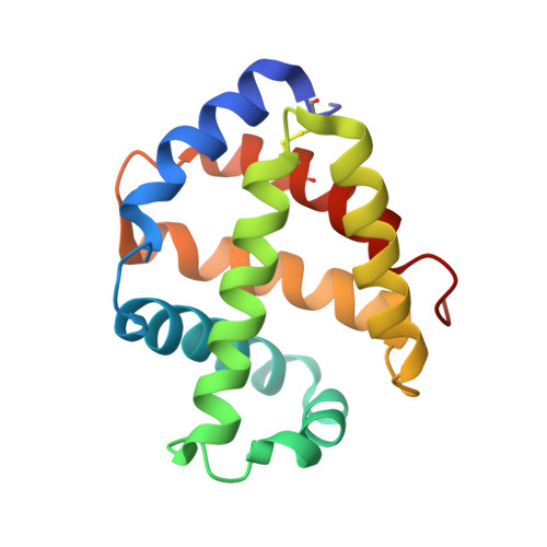
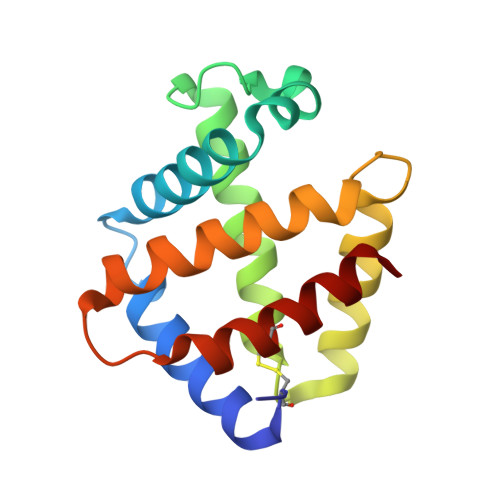
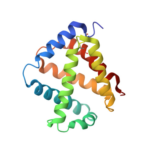
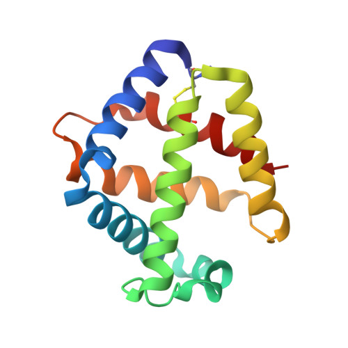
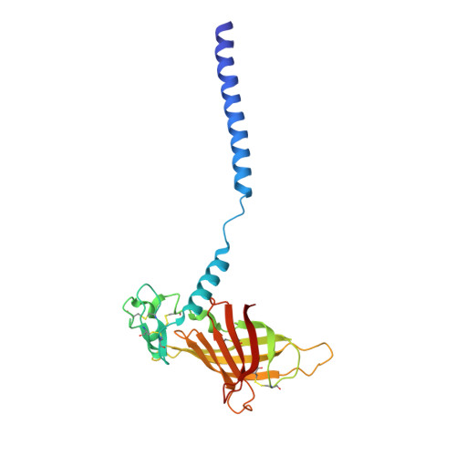
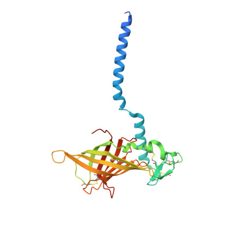
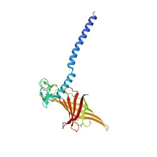
The structure of the giant haemoglobin from Glossoscolex paulistus.

Ruggiero Bachega, J.F.Vasconcelos Maluf, F.Andi, B.D'Muniz Pereira, H.Falsarella Carazzollea, M.Orville, A.M.Tabak, M.Brandao-Neto, J.Garratt, R.C.Horjales Reboredo, E.

(2015) Acta Crystallogr D Biol Crystallogr 71: 1257-1271

  • DOI: https://doi.org/10.1107/S1399004715005453
  • Primary Citation of Related Structures:  
    4U8U, 4WCH

  • PubMed Abstract: 

    The sequences of all seven polypeptide chains from the giant haemoglobin of the free-living earthworm Glossoscolex paulistus (HbGp) are reported together with the three-dimensional structure of the 3.6 MDa complex which they form. The refinement of the full particle, which has been solved at 3.2 Å resolution, the highest resolution reported to date for a hexagonal bilayer haemoglobin composed of 12 protomers, is reported. This has allowed a more detailed description of the contacts between subunits which are essential for particle stability. Interpretation of features in the electron-density maps suggests the presence of metal-binding sites (probably Zn(2+) and Ca(2+)) and glycosylation sites, some of which have not been reported previously. The former appear to be important for the integrity of the particle. The crystal structure of the isolated d chain (d-HbGp) at 2.1 Å resolution shows different interchain contacts between d monomers compared with those observed in the full particle. Instead of forming trimers, as seen in the complex, the isolated d chains associate to form dimers across a crystallographic twofold axis. These observations eliminate the possibility that trimers form spontaneously in solution as intermediates during the formation of the dodecameric globin cap and contribute to understanding of the possible ways in which the particle self-assembles.


  • Organizational Affiliation

    Instituto de Física de São Carlos, Universidade de São Paulo, São Carlos, Brazil.


Macromolecules
Find similar proteins by:  (by identity cutoff)  |  3D Structure
Entity ID: 1
MoleculeChains Sequence LengthOrganismDetailsImage
Globin a chain150Glossoscolex paulistusMutation(s): 0 
UniProt
Find proteins for A0A0M3KKX5 (Glossoscolex paulistus)
Explore A0A0M3KKX5 
Go to UniProtKB:  A0A0M3KKX5
Entity Groups  
Sequence Clusters30% Identity50% Identity70% Identity90% Identity95% Identity100% Identity
UniProt GroupA0A0M3KKX5
Sequence Annotations
Expand
  • Reference Sequence
Find similar proteins by:  (by identity cutoff)  |  3D Structure
Entity ID: 2
MoleculeChains Sequence LengthOrganismDetailsImage
Globin b Chain142Glossoscolex paulistusMutation(s): 0 
UniProt
Find proteins for A0A0M3KKX6 (Glossoscolex paulistus)
Explore A0A0M3KKX6 
Go to UniProtKB:  A0A0M3KKX6
Entity Groups  
Sequence Clusters30% Identity50% Identity70% Identity90% Identity95% Identity100% Identity
UniProt GroupA0A0M3KKX6
Sequence Annotations
Expand
  • Reference Sequence
Find similar proteins by:  (by identity cutoff)  |  3D Structure
Entity ID: 3
MoleculeChains Sequence LengthOrganismDetailsImage
Globin c Chain151Glossoscolex paulistusMutation(s): 0 
UniProt
Find proteins for A0A0M3KKX7 (Glossoscolex paulistus)
Explore A0A0M3KKX7 
Go to UniProtKB:  A0A0M3KKX7
Entity Groups  
Sequence Clusters30% Identity50% Identity70% Identity90% Identity95% Identity100% Identity
UniProt GroupA0A0M3KKX7
Sequence Annotations
Expand
  • Reference Sequence
Find similar proteins by:  (by identity cutoff)  |  3D Structure
Entity ID: 4
MoleculeChains Sequence LengthOrganismDetailsImage
Globin d Chain141Glossoscolex paulistusMutation(s): 0 
UniProt
Find proteins for A0A0M3KKX8 (Glossoscolex paulistus)
Explore A0A0M3KKX8 
Go to UniProtKB:  A0A0M3KKX8
Entity Groups  
Sequence Clusters30% Identity50% Identity70% Identity90% Identity95% Identity100% Identity
UniProt GroupA0A0M3KKX8
Sequence Annotations
Expand
  • Reference Sequence
Find similar proteins by:  (by identity cutoff)  |  3D Structure
Entity ID: 5
MoleculeChains Sequence LengthOrganismDetailsImage
Linker L1BA [auth b],
M,
QA [auth q]
224Glossoscolex paulistusMutation(s): 0 
UniProt
Find proteins for A0A0M3KKX9 (Glossoscolex paulistus)
Explore A0A0M3KKX9 
Go to UniProtKB:  A0A0M3KKX9
Entity Groups  
Sequence Clusters30% Identity50% Identity70% Identity90% Identity95% Identity100% Identity
UniProt GroupA0A0M3KKX9
Sequence Annotations
Expand
  • Reference Sequence
Find similar proteins by:  (by identity cutoff)  |  3D Structure
Entity ID: 6
MoleculeChains Sequence LengthOrganismDetailsImage
Linker L2CA [auth c],
N,
RA [auth r]
236Glossoscolex paulistusMutation(s): 0 
UniProt
Find proteins for A0A0M3KKY0 (Glossoscolex paulistus)
Explore A0A0M3KKY0 
Go to UniProtKB:  A0A0M3KKY0
Entity Groups  
Sequence Clusters30% Identity50% Identity70% Identity90% Identity95% Identity100% Identity
UniProt GroupA0A0M3KKY0
Sequence Annotations
Expand
  • Reference Sequence
Find similar proteins by:  (by identity cutoff)  |  3D Structure
Entity ID: 7
MoleculeChains Sequence LengthOrganismDetailsImage
Linker L3DA [auth d],
O,
SA [auth s]
218Glossoscolex paulistusMutation(s): 0 
UniProt
Find proteins for A0A0M3KKY1 (Glossoscolex paulistus)
Explore A0A0M3KKY1 
Go to UniProtKB:  A0A0M3KKY1
Entity Groups  
Sequence Clusters30% Identity50% Identity70% Identity90% Identity95% Identity100% Identity
UniProt GroupA0A0M3KKY1
Sequence Annotations
Expand
  • Reference Sequence
Small Molecules
Ligands 5 Unique
IDChains Name / Formula / InChI Key2D Diagram3D Interactions
HEM
Query on HEM

Download Ideal Coordinates CCD File 
BB [auth E]
BC [auth R]
CD [auth e]
DB [auth F]
DC [auth S]
BB [auth E],
BC [auth R],
CD [auth e],
DB [auth F],
DC [auth S],
ED [auth f],
FB [auth G],
FC [auth T],
GD [auth g],
HB [auth H],
HC [auth U],
ID [auth h],
JB [auth I],
JC [auth V],
KD [auth i],
LB [auth J],
LC [auth W],
MD [auth j],
NB [auth K],
NC [auth X],
OD [auth k],
PB [auth L],
PC [auth Y],
QD [auth l],
RC [auth Z],
SD [auth m],
TA [auth A],
TC [auth a],
UD [auth n],
VA [auth B],
WD [auth o],
XA [auth C],
XB [auth P],
YD [auth p],
ZA [auth D],
ZB [auth Q]
PROTOPORPHYRIN IX CONTAINING FE
C34 H32 Fe N4 O4
KABFMIBPWCXCRK-RGGAHWMASA-L
NAG
Query on NAG

Download Ideal Coordinates CCD File 
BD [auth d],
EE [auth s],
WB [auth O]
2-acetamido-2-deoxy-beta-D-glucopyranose
C8 H15 N O6
OVRNDRQMDRJTHS-FMDGEEDCSA-N
ZN
Query on ZN

Download Ideal Coordinates CCD File 
AD [auth d]
DE [auth s]
UB [auth O]
VB [auth O]
XC [auth c]
AD [auth d],
DE [auth s],
UB [auth O],
VB [auth O],
XC [auth c],
ZC [auth d]
ZINC ION
Zn
PTFCDOFLOPIGGS-UHFFFAOYSA-N
CA
Query on CA

Download Ideal Coordinates CCD File 
AE [auth q]
BE [auth r]
CE [auth s]
RB [auth M]
SB [auth N]
AE [auth q],
BE [auth r],
CE [auth s],
RB [auth M],
SB [auth N],
TB [auth O],
VC [auth b],
WC [auth c],
YC [auth d]
CALCIUM ION
Ca
BHPQYMZQTOCNFJ-UHFFFAOYSA-N
CYN
Query on CYN

Download Ideal Coordinates CCD File 
AB [auth D]
AC [auth Q]
CB [auth E]
CC [auth R]
DD [auth e]
AB [auth D],
AC [auth Q],
CB [auth E],
CC [auth R],
DD [auth e],
EB [auth F],
EC [auth S],
FD [auth f],
GB [auth G],
GC [auth T],
HD [auth g],
IB [auth H],
IC [auth U],
JD [auth h],
KB [auth I],
KC [auth V],
LD [auth i],
MB [auth J],
MC [auth W],
ND [auth j],
OB [auth K],
OC [auth X],
PD [auth k],
QB [auth L],
QC [auth Y],
RD [auth l],
SC [auth Z],
TD [auth m],
UA [auth A],
UC [auth a],
VD [auth n],
WA [auth B],
XD [auth o],
YA [auth C],
YB [auth P],
ZD [auth p]
CYANIDE ION
C N
XFXPMWWXUTWYJX-UHFFFAOYSA-N
Experimental Data & Validation

Experimental Data

  • Method: X-RAY DIFFRACTION
  • Resolution: 3.20 Å
  • R-Value Free: 0.235 
  • R-Value Work: 0.215 
  • R-Value Observed: 0.216 
  • Space Group: I 2 2 2
Unit Cell:
Length ( Å )Angle ( ˚ )
a = 272.68α = 90
b = 319.9β = 90
c = 333.18γ = 90
Software Package:
Software NamePurpose
PHENIXrefinement

Structure Validation

View Full Validation Report


Ligand Structure Quality Assessment 


Entry History 

Deposition Data

Revision History  (Full details and data files)

  • Version 1.0: 2015-06-10
    Type: Initial release
  • Version 1.1: 2015-06-17
    Changes: Structure summary
  • Version 1.2: 2015-06-24
    Changes: Database references
  • Version 1.3: 2016-09-07
    Changes: Derived calculations, Non-polymer description
  • Version 1.4: 2020-07-29
    Type: Remediation
    Reason: Carbohydrate remediation
    Changes: Data collection, Derived calculations, Structure summary
  • Version 1.5: 2023-12-27
    Changes: Data collection, Database references, Structure summary