Structure based design of novel 6,5 heterobicyclic mitogen-activated protein kinase kinase (MEK) inhibitors leading to the discovery of imidazo[1,5-a] pyrazine G-479.
Robarge, K.D., Lee, W., Eigenbrot, C., Ultsch, M., Wiesmann, C., Heald, R., Price, S., Hewitt, J., Jackson, P., Savy, P., Burton, B., Choo, E.F., Pang, J., Boggs, J., Yang, A., Yang, X., Baumgardner, M.(2014) Bioorg Med Chem Lett 24: 4714-4723
- PubMed: 25193232
- DOI: https://doi.org/10.1016/j.bmcl.2014.08.008
- Primary Citation of Related Structures:
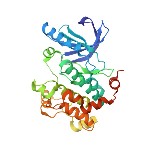
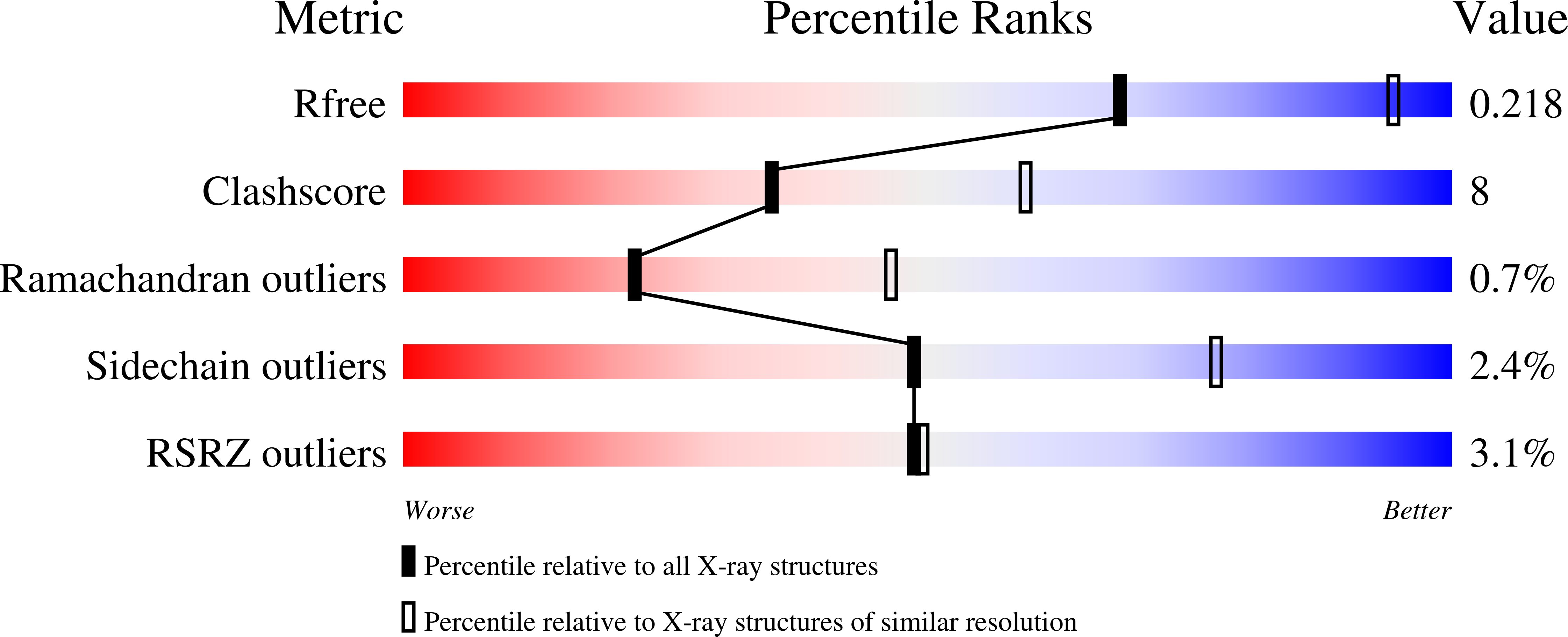
4U7Z, 4U80, 4U81 - PubMed Abstract:
Use of the tools of SBDD including crystallography led to the discovery of novel and potent 6,5 heterobicyclic MEKi's [J. Med. Chem.2012, 55, 4594]. The core change from a 5,6 heterobicycle to a 6,5 heterobicycle was driven by the desire for increased structural diversity and aided by the co-crystal structure of G-925 [J. Med. Chem.2012, 55, 4594]. The key design feature was the shift of the attachment of the five-membered heterocyclic ring towards the B ring while maintaining the key hydroxamate and anilino pharamcophoric elements in a remarkably similar position as in G-925. From modelling, changing the connection point of the five membered ring heterocycle placed the H-bond accepting nitrogen within a good distance and angle to the Ser212 [J. Med. Chem.2012, 55, 4594]. The resulting novel 6,5 benzoisothiazole MEKi G-155 exhibited improved potency versus aza-benzofurans G-925 and G-963 but was a potent inhibitor of cytochrome P450's 2C9 and 2C19. Lowering the logD by switching to the more polar imidazo[1,5-a] pyridine core significantly diminished 2C9/2C19 inhibition while retaining potency. The imidazo[1,5-a] pyridine G-868 exhibited increased potency versus the starting point for this work (aza-benzofuran G-925) leading to deprioritization of the azabenzofurans. The 6,5-imidazo[1,5-a] pyridine scaffold was further diversified by incorporating a nitrogen at the 7 position to give the imidazo[1,5-a] pyrazine scaffold. The introduction of the C7 nitrogen was driven by the desire to improve metabolic stability by blocking metabolism at the C7 and C8 positions (particularly the HLM stability). It was found that improving on G-868 (later renamed GDC-0623) required combining C7 nitrogen with a diol hydroxamate to give G-479. G-479 with polarity distributed throughout the molecule was improved over G-868 in many aspects.
Organizational Affiliation:
Discovery Chemistry, Genentech Inc., 1 DNA Way, South San Francisco, CA, USA. Electronic address: kir@gene.com.