4U5J

C-Src in complex with Ruxolitinib


Experimental Data Snapshot

  • Method: X-RAY DIFFRACTION
  • Resolution: 2.26 Å
  • R-Value Free: 0.236 
  • R-Value Work: 0.197 
  • R-Value Observed: 0.199 

wwPDB Validation   3D Report Full Report


Ligand Structure Quality Assessment 


This is version 1.2 of the entry. See complete history


Literature

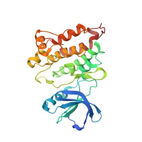
c-Src Binds to the Cancer Drug Ruxolitinib with an Active Conformation

Duan, Y.Chen, L.Chen, Y.Fan, X.G.

(2014) PLoS One 9: e106225-e106225

  • DOI: https://doi.org/10.1371/journal.pone.0106225
  • Primary Citation of Related Structures:  
    4U5J

  • PubMed Abstract: 

    The cancer drug Ruxolitinib is a potent janus kinase inhibitor approved for the treatment of the myeloproliferative neoplasms. In addition, Ruxolitinib has weak inhibitory activity against a panel of other kinases, including Src kinase. There is no structural information of Ruxolitinib binding to any kinase. In this paper, we determined the crystal structure of c-Src kinase domain in complex of Ruxolitinib at a resolution of 2.26 Å. C-Src kinase domain adopts the DFG-in active conformation upon Ruxolitinib binding, indicating Ruxolitinib is a type I inhibitor for c-Src. Ruxolitinib forms two hydrogen bonds with Met341, a water-mediated hydrogen bond with Thr338, and a number of van der Waals contacts with c-Src. Ruxolitinib was then docked into the ligand-binding pocket of a previously solved JAK1 structure. From the docking result, Ruxolitinib also binds JAK1 as a type I inhibitor, with more interactions and a higher shape complementarity with the ligand-binding pocket of JAK1 compared to that of c-Src. Since Ruxolitinib is a relatively small inhibitor and there is sizeable cavity between Ruxolitinib and c-Src ligand-binding pocket, we propose to modify Ruxolitinib to develop more potent inhibitors to c-Src.


  • Organizational Affiliation

    Department of Infectious Diseases & Laboratory of Structural Biology, Key Laboratory of Cancer Proteomics of Chinese Ministry of Health, XiangYa Hospital, Central South University, Changsha, Hunan, China.


Macromolecules
Find similar proteins by:  (by identity cutoff)  |  3D Structure
Entity ID: 1
MoleculeChains Sequence LengthOrganismDetailsImage
Proto-oncogene tyrosine-protein kinase Src
A, B
286Gallus gallusMutation(s): 0 
Gene Names: SRC
EC: 2.7.10.2
UniProt
Find proteins for P00523 (Gallus gallus)
Explore P00523 
Go to UniProtKB:  P00523
Entity Groups  
Sequence Clusters30% Identity50% Identity70% Identity90% Identity95% Identity100% Identity
UniProt GroupP00523
Sequence Annotations
Expand
  • Reference Sequence
Small Molecules
Ligands 1 Unique
IDChains Name / Formula / InChI Key2D Diagram3D Interactions
RXT
Query on RXT

Download Ideal Coordinates CCD File 
C [auth A],
D [auth B]
(3R)-3-cyclopentyl-3-[4-(7H-pyrrolo[2,3-d]pyrimidin-4-yl)-1H-pyrazol-1-yl]propanenitrile
C17 H18 N6
HFNKQEVNSGCOJV-OAHLLOKOSA-N
Binding Affinity Annotations 
IDSourceBinding Affinity
RXT BindingDB:  4U5J Kd: min: 1.00e+4, max: 1.00e+4 (nM) from 2 assay(s)
Binding MOAD:  4U5J IC50: 2920 (nM) from 1 assay(s)
Experimental Data & Validation

Experimental Data

  • Method: X-RAY DIFFRACTION
  • Resolution: 2.26 Å
  • R-Value Free: 0.236 
  • R-Value Work: 0.197 
  • R-Value Observed: 0.199 
  • Space Group: P 1
Unit Cell:
Length ( Å )Angle ( ˚ )
a = 42.107α = 79.27
b = 63.228β = 89.27
c = 73.989γ = 90.29
Software Package:
Software NamePurpose
PHENIXrefinement

Structure Validation

View Full Validation Report


Ligand Structure Quality Assessment 


Entry History & Funding Information

Deposition Data


Funding OrganizationLocationGrant Number
National Natural Science Foundation of ChinaChina81272971
National Natural Science Foundation of ChinaChina81372904
National Institutes of Health/National Institute of General Medical Sciences (NIH/NIGMS)United States5R01GM064642

Revision History  (Full details and data files)

  • Version 1.0: 2014-09-17
    Type: Initial release
  • Version 1.1: 2017-10-18
    Changes: Advisory, Author supporting evidence, Derived calculations, Other, Source and taxonomy, Structure summary
  • Version 1.2: 2022-03-23
    Changes: Author supporting evidence, Database references, Refinement description