4U0S

Structure of Eukaryotic fic domain containing protein with ADP


Experimental Data Snapshot

  • Method: X-RAY DIFFRACTION
  • Resolution: 2.49 Å
  • R-Value Free: 0.245 
  • R-Value Work: 0.207 
  • R-Value Observed: 0.209 

wwPDB Validation   3D Report Full Report


Ligand Structure Quality Assessment 


This is version 1.2 of the entry. See complete history


Literature

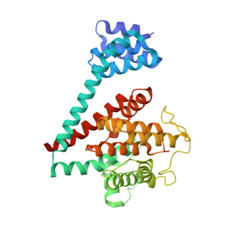
Crystal structure of the human, FIC-domain containing protein HYPE and implications for its functions.

Bunney, T.D.Cole, A.R.Broncel, M.Esposito, D.Tate, E.W.Katan, M.

(2014) Structure 22: 1831-1843

  • DOI: https://doi.org/10.1016/j.str.2014.10.007
  • Primary Citation of Related Structures:  
    4U04, 4U07, 4U0S, 4U0U, 4U0Z

  • PubMed Abstract: 

    Protein AMPylation, the transfer of AMP from ATP to protein targets, has been recognized as a new mechanism of host-cell disruption by some bacterial effectors that typically contain a FIC-domain. Eukaryotic genomes also encode one FIC-domain protein,HYPE, which has remained poorly characterized.Here we describe the structure of human HYPE, solved by X-ray crystallography, representing the first structure of a eukaryotic FIC-domain protein. We demonstrate that HYPE forms stable dimers with structurally and functionally integrated FIC-domains and with TPR-motifs exposed for protein-protein interactions. As HYPE also uniquely possesses a transmembrane helix, dimerization is likely to affect its positioning and function in the membrane vicinity. The low rate of auto AMPylation of the wild-type HYPE could be due to autoinhibition, consistent with the mechanism proposed for a number of putative FIC AMPylators. Our findings also provide a basis to further consider possible alternative cofactors of HYPE and distinct modes of target-recognition.


  • Organizational Affiliation

    Division of Biosciences, Institute of Structural and Molecular Biology, University College London, Gower Street, London WC1E 6BT, UK. Electronic address: t.bunney@ucl.ac.uk.


Macromolecules
Find similar proteins by:  (by identity cutoff)  |  3D Structure
Entity ID: 1
MoleculeChains Sequence LengthOrganismDetailsImage
Adenosine monophosphate-protein transferase FICD
A, B
344Homo sapiensMutation(s): 1 
Gene Names: FICDHIP13HYPEUNQ3041/PRO9857
EC: 2.7.7
UniProt & NIH Common Fund Data Resources
Find proteins for Q9BVA6 (Homo sapiens)
Explore Q9BVA6 
Go to UniProtKB:  Q9BVA6
PHAROS:  Q9BVA6
GTEx:  ENSG00000198855 
Entity Groups  
Sequence Clusters30% Identity50% Identity70% Identity90% Identity95% Identity100% Identity
UniProt GroupQ9BVA6
Sequence Annotations
Expand
  • Reference Sequence
Experimental Data & Validation

Experimental Data

  • Method: X-RAY DIFFRACTION
  • Resolution: 2.49 Å
  • R-Value Free: 0.245 
  • R-Value Work: 0.207 
  • R-Value Observed: 0.209 
  • Space Group: P 1 21 1
Unit Cell:
Length ( Å )Angle ( ˚ )
a = 71.04α = 90
b = 76.01β = 107.56
c = 92.03γ = 90
Software Package:
Software NamePurpose
MOSFLMdata reduction
Aimlessdata scaling
PHASERphasing
Cootmodel building
BUSTERrefinement

Structure Validation

View Full Validation Report


Ligand Structure Quality Assessment 


Entry History 

Deposition Data

Revision History  (Full details and data files)

  • Version 1.0: 2014-12-10
    Type: Initial release
  • Version 1.1: 2015-02-04
    Changes: Database references
  • Version 1.2: 2023-12-20
    Changes: Data collection, Database references, Derived calculations, Refinement description