4TWO

Human EphA3 Kinase domain in complex with compound 164


Experimental Data Snapshot

  • Method: X-RAY DIFFRACTION
  • Resolution: 2.05 Å
  • R-Value Free: 0.201 
  • R-Value Work: 0.163 
  • R-Value Observed: 0.165 

wwPDB Validation   3D Report Full Report


Ligand Structure Quality Assessment 


This is version 1.1 of the entry. See complete history


Literature

Structural Analysis of the Binding of Type I, I1/2, and II Inhibitors to Eph Tyrosine Kinases.

Dong, J.Zhao, H.Zhou, T.Spiliotopoulos, D.Rajendran, C.Li, X.D.Huang, D.Caflisch, A.

(2015) ACS Med Chem Lett 6: 79-83

  • DOI: https://doi.org/10.1021/ml500355x
  • Primary Citation of Related Structures:  
    4TWN, 4TWO

  • PubMed Abstract: 

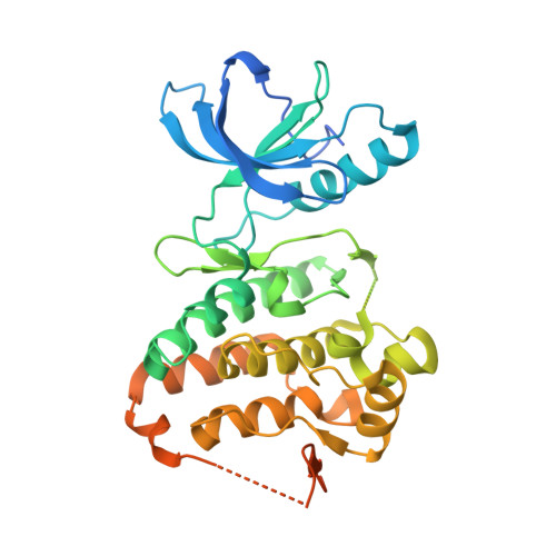
    We have solved the crystal structures of the EphA3 tyrosine kinase in complex with nine small-molecule inhibitors, which represent five different chemotypes and three main binding modes, i.e., types I and I1/2 (DFG in) and type II (DFG out). The three structures with type I1/2 inhibitors show that the higher affinity with respect to type I is due to an additional polar group (hydroxyl or pyrazole ring of indazole) which is fully buried and is involved in the same hydrogen bonds as the (urea or amide) linker of the type II inhibitors. Overall, the type I and type II binding modes belong to the lock-and-key and induced fit mechanism, respectively. In the type II binding, the scaffold in contact with the hinge region influences the position of the Phe765 side chain of the DFG motif and the orientation of the Gly-rich loop. The binding mode of Birb796 in the EphA3 kinase does not involve any hydrogen bond with the hinge region, which is different from the Birb796/p38 MAP kinase complex. Our structural analysis emphasizes the importance of accounting for structural plasticity of the ATP binding site in the design of type II inhibitors of tyrosine kinases.


  • Organizational Affiliation

    Department of Biochemistry, University of Zurich , Winterthurerstrasse 190, CH-8057 Zurich, Switzerland.


Macromolecules
Find similar proteins by:  (by identity cutoff)  |  3D Structure
Entity ID: 1
MoleculeChains Sequence LengthOrganismDetailsImage
Ephrin type-A receptor 3361Homo sapiensMutation(s): 0 
Gene Names: EPHA3ETKETK1HEKTYRO4
EC: 2.7.10.1
UniProt & NIH Common Fund Data Resources
Find proteins for P29320 (Homo sapiens)
Explore P29320 
Go to UniProtKB:  P29320
PHAROS:  P29320
GTEx:  ENSG00000044524 
Entity Groups  
Sequence Clusters30% Identity50% Identity70% Identity90% Identity95% Identity100% Identity
UniProt GroupP29320
Sequence Annotations
Expand
  • Reference Sequence
Small Molecules
Ligands 1 Unique
IDChains Name / Formula / InChI Key2D Diagram3D Interactions
37W
Query on 37W

Download Ideal Coordinates CCD File 
B [auth A]5-{[3-carbamoyl-4-(3,4-dimethylphenyl)-5-methylthiophen-2-yl]amino}-5-oxopentanoic acid
C19 H22 N2 O4 S
ZOPDRGUNCASWKP-UHFFFAOYSA-N
Binding Affinity Annotations 
IDSourceBinding Affinity
37W Binding MOAD:  4TWO IC50: 8000 (nM) from 1 assay(s)
Experimental Data & Validation

Experimental Data

  • Method: X-RAY DIFFRACTION
  • Resolution: 2.05 Å
  • R-Value Free: 0.201 
  • R-Value Work: 0.163 
  • R-Value Observed: 0.165 
  • Space Group: P 1 21 1
Unit Cell:
Length ( Å )Angle ( ˚ )
a = 53.221α = 90
b = 38.068β = 100.83
c = 75.31γ = 90
Software Package:
Software NamePurpose
PHENIXrefinement
SCALAdata scaling
XDSdata reduction
Cootmodel building
PDB_EXTRACTdata extraction
PHASERphasing
XSCALEdata reduction

Structure Validation

View Full Validation Report


Ligand Structure Quality Assessment 


Entry History 

Deposition Data

Revision History  (Full details and data files)

  • Version 1.0: 2015-05-13
    Type: Initial release
  • Version 1.1: 2023-12-20
    Changes: Data collection, Database references, Refinement description