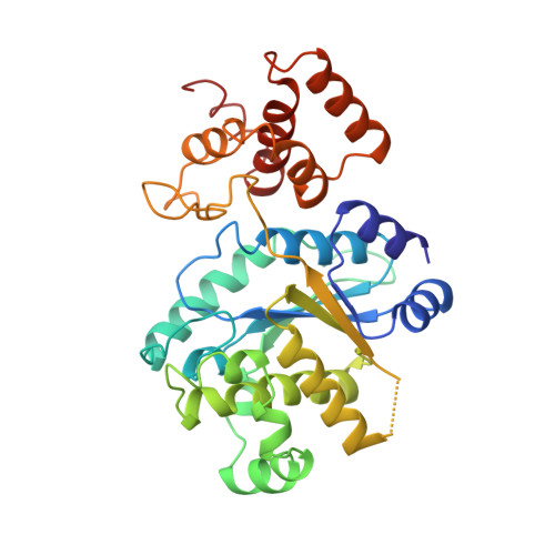
Crystal structure of a deletion mutant of a tyrosyl-tRNA synthetase complexed with tyrosine.
Brick, P., Blow, D.M.(1987) J Mol Biol 194: 287-297
- PubMed: 3612807
- DOI: https://doi.org/10.1016/0022-2836(87)90376-7
- Primary Citation of Related Structures:
4TS1 - PubMed Abstract:
The crystal structure of a deletion mutant of tyrosyl-tRNA synthetase from Bacillus stearothermophilus has been determined at 2.5 A resolution using molecular replacement techniques. The genetically engineered molecule catalyses the activation of tyrosine with kinetic properties similar to those of the wild-type enzyme but no longer binds tRNATyr. It contains 319 residues corresponding to the region of the polypeptide chain for which interpretable electron density is present in crystals of the wild-type enzyme. The partly refined model of the wild-type enzyme was used as a starting point in determining the structure of the truncated mutant. The new crystals are of space group P2(1) and contain the molecular dimer within the asymmetric unit. The refined model has a crystallographic R-factor of 18.7% for all reflections between 8 and 2.5 A. Each subunit contains two structural domains: the alpha/beta domain (residues 1 to 220) containing a six-stranded beta-sheet and the alpha-helical domain (residues 248 to 319) containing five helices. The alpha/beta domains are related by a non-crystallographic dyad while the alpha-helical domains are in slightly different orientations in the two subunits. The tyrosine substrate binds in a slot at the bottom of a deep active site cleft in the middle of the alpha/beta domain. It is surrounded by polar side-chains and water molecules that are involved in an intricate hydrogen bonding network. Both the alpha-amino and hydroxyl groups of the substrate make good hydrogen bonds with the protein. The amino group forms hydrogen bonds with Tyr169-OH, Asp78-OD1 and Gln173-OE1. The phenolic hydroxyl group forms hydrogen bonds with Asp76-OD1 and Tyr34-OH. In contrast, the substrate carboxyl group makes no direct interactions with the enzyme. The results of both substrate inhibition studies and site-directed mutagenesis experiments have been examined in the light of the refined structure.