4TR1

Crystal structure of GSH-bound cGrx2/C15S


Experimental Data Snapshot

  • Method: X-RAY DIFFRACTION
  • Resolution: 1.58 Å
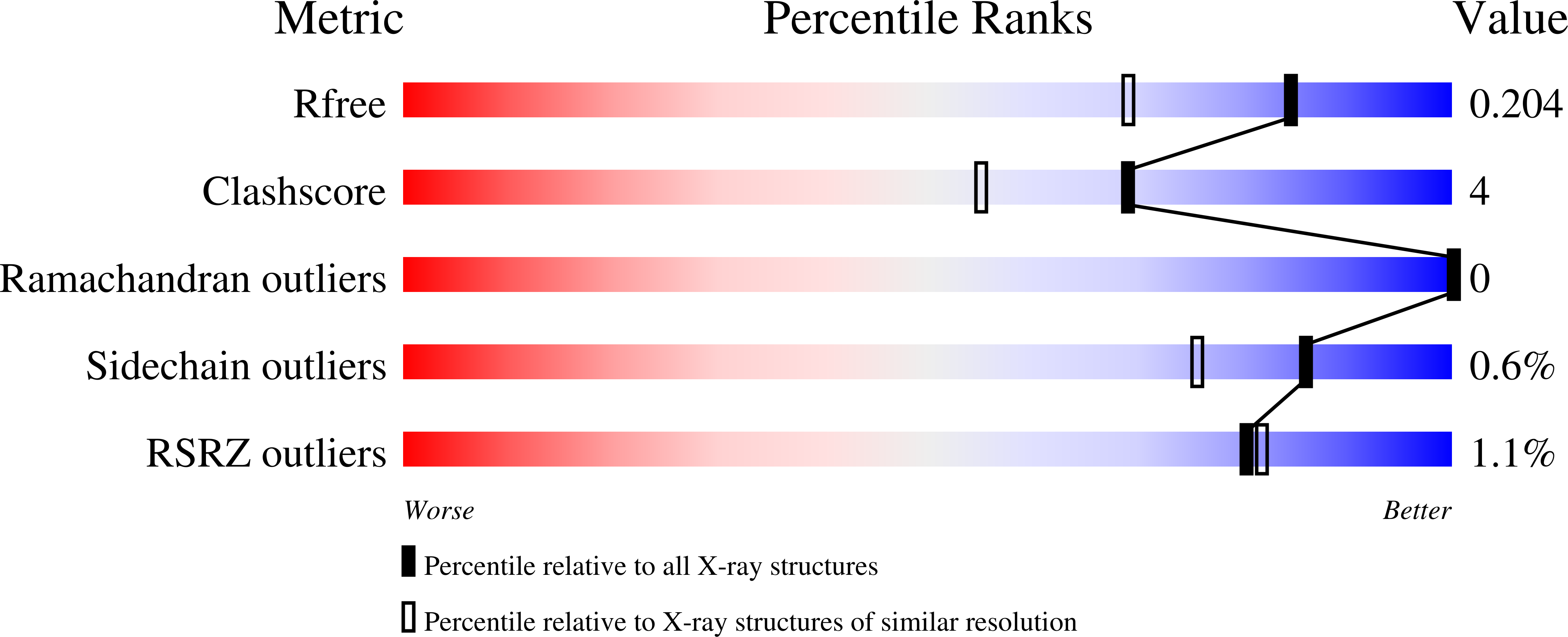
  • R-Value Free: 0.204 
  • R-Value Work: 0.177 
  • R-Value Observed: 0.179 

wwPDB Validation   3D Report Full Report


Ligand Structure Quality Assessment 


This is version 1.5 of the entry. See complete history


Literature

The GSH- and GSSG-bound structures of glutaredoxin from Clostridium oremlandii.

Lee, E.H.Kim, H.Y.Hwang, K.Y.

(2014) Arch Biochem Biophys 564C: 20-25

  • DOI: https://doi.org/10.1016/j.abb.2014.09.001
  • Primary Citation of Related Structures:  
    4TR0, 4TR1

  • PubMed Abstract: 

    Glutaredoxin (Grx) is a major redox enzyme that reduces disulfide bonds using glutathione (GSH) as an electron donor. The anaerobic bacterium Clostridium oremlandii possesses a selenocysteine-containing Grx (cGrx1) and a cysteine-containing homolog (cGrx2). Here, the crystal structure of the GSSG-bound form of cGrx2 was determined for the first time at a resolution of 1.95Å. In addition, its monothiol variant cGrx2/C15S in complex with GSH was also determined at a resolution of 1.58Å. cGrx2 is a monomeric protein with an overall structure that consists of the typical thioredoxin fold composed of four α-helices and four β-strands. Two ligands, GSH and GSSG, share a conserved binding site consisting of CPYC, TVP, and CDD motifs. The cysteinyl and γ-glutamyl moieties show similar binding interactions in the two structures, whereas the glycine moiety shows different interactions. Interestingly, the structures revealed that only one GSH moiety of GSSG is sufficient for its binding to the protein. The GSSG-bound structure of cGrx2 was obtained as an oxidized form with a disulfide bond at the CPYC motif. Comparison of the GSH-binding mode in cGrx2 to other known Grxs revealed similarities as well as some diversity.


  • Organizational Affiliation

    Division of Biotechnology, College of Life Sciences and Biotechnology, Korea University, Seoul, Republic of Korea.


Macromolecules
Find similar proteins by:  (by identity cutoff)  |  3D Structure
Entity ID: 1
MoleculeChains Sequence LengthOrganismDetailsImage
Glutaredoxin 392Alkaliphilus oremlandii OhILAsMutation(s): 1 
Gene Names: Clos_2422
UniProt
Find proteins for A8MJH2 (Alkaliphilus oremlandii (strain OhILAs))
Explore A8MJH2 
Go to UniProtKB:  A8MJH2
Entity Groups  
Sequence Clusters30% Identity50% Identity70% Identity90% Identity95% Identity100% Identity
UniProt GroupA8MJH2
Sequence Annotations
Expand
  • Reference Sequence
Find similar proteins by:  (by identity cutoff)  |  3D Structure
Entity ID: 2
MoleculeChains Sequence LengthOrganismDetailsImage
Glutaredoxin 392Alkaliphilus oremlandii OhILAsMutation(s): 1 
Gene Names: Clos_2422
UniProt
Find proteins for A8MJH2 (Alkaliphilus oremlandii (strain OhILAs))
Explore A8MJH2 
Go to UniProtKB:  A8MJH2
Entity Groups  
Sequence Clusters30% Identity50% Identity70% Identity90% Identity95% Identity100% Identity
UniProt GroupA8MJH2
Sequence Annotations
Expand
  • Reference Sequence
Small Molecules
Modified Residues  2 Unique
IDChains TypeFormula2D DiagramParent
CSO
Query on CSO
A
L-PEPTIDE LINKINGC3 H7 N O3 SCYS
CSU
Query on CSU
B
L-PEPTIDE LINKINGC3 H7 N O5 S2CYS
Experimental Data & Validation

Experimental Data

  • Method: X-RAY DIFFRACTION
  • Resolution: 1.58 Å
  • R-Value Free: 0.204 
  • R-Value Work: 0.177 
  • R-Value Observed: 0.179 
  • Space Group: C 1 2 1
Unit Cell:
Length ( Å )Angle ( ˚ )
a = 123.065α = 90
b = 29.371β = 118.17
c = 64.836γ = 90
Software Package:
Software NamePurpose
HKL-2000data scaling
PDB_EXTRACTdata extraction
PHENIXrefinement

Structure Validation

View Full Validation Report


Ligand Structure Quality Assessment 


Entry History 

Deposition Data

Revision History  (Full details and data files)

  • Version 1.0: 2014-10-01
    Type: Initial release
  • Version 1.1: 2014-12-17
    Changes: Database references
  • Version 1.2: 2015-01-07
    Changes: Database references
  • Version 1.3: 2015-02-04
    Changes: Derived calculations
  • Version 1.4: 2017-11-22
    Changes: Advisory, Database references, Derived calculations, Other, Refinement description, Source and taxonomy
  • Version 1.5: 2023-12-27
    Changes: Data collection, Database references, Refinement description