4TPG

Selectivity mechanism of a bacterial homologue of the human drug peptide transporters PepT1 and PepT2


Experimental Data Snapshot

  • Method: X-RAY DIFFRACTION
  • Resolution: 3.91 Å
  • R-Value Free: 0.290 
  • R-Value Work: 0.261 
  • R-Value Observed: 0.262 

wwPDB Validation   3D Report Full Report


Ligand Structure Quality Assessment 


This is version 1.4 of the entry. See complete history


Literature

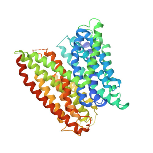
Selectivity mechanism of a bacterial homolog of the human drug-peptide transporters PepT1 and PepT2.

Guettou, F.Quistgaard, E.M.Raba, M.Moberg, P.Low, C.Nordlund, P.

(2014) Nat Struct Mol Biol 21: 728

  • DOI: https://doi.org/10.1038/nsmb.2860
  • Primary Citation of Related Structures:  
    4TPG, 4TPH, 4TPJ

  • PubMed Abstract: 

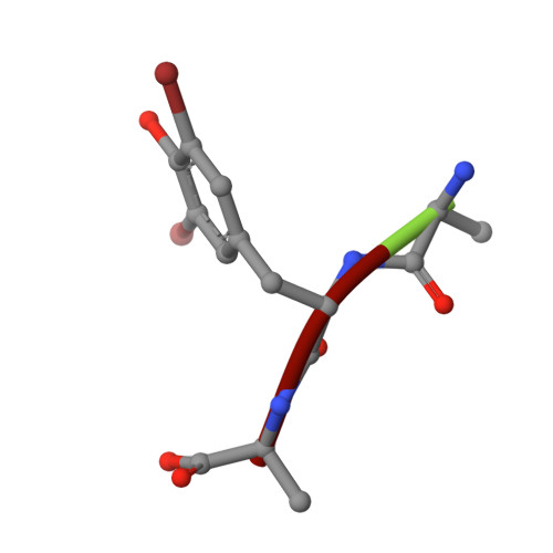
    Peptide transporters of the PepT family have key roles in the transport of di- and tripeptides across membranes as well as in the absorption of orally administered drugs in the small intestine. We have determined structures of a PepT transporter from Shewanella oneidensis (PepT(So2)) in complex with three different peptides. The peptides bind in a large cavity lined by residues that are highly conserved in human PepT1 and PepT2. The bound peptides adopt extended conformations with their N termini clamped into a conserved polar pocket. A positively charged patch allows differential interactions with the C-terminal carboxylates of di- and tripeptides. Here we identify three pockets for peptide side chain interactions, and our binding studies define differential roles of these pockets for the recognition of different subtypes of peptide side chains.


  • Organizational Affiliation

    Department of Medical Biochemistry and Biophysics, Karolinska Institutet, Stockholm, Sweden.


Macromolecules
Find similar proteins by:  (by identity cutoff)  |  3D Structure
Entity ID: 1
MoleculeChains Sequence LengthOrganismDetailsImage
Proton:oligopeptide symporter POT family
A, B
523Shewanella oneidensis MR-1Mutation(s): 0 
Gene Names: SO_1277
Membrane Entity: Yes 
UniProt
Find proteins for Q8EHE6 (Shewanella oneidensis (strain MR-1))
Explore Q8EHE6 
Go to UniProtKB:  Q8EHE6
Entity Groups  
Sequence Clusters30% Identity50% Identity70% Identity90% Identity95% Identity100% Identity
UniProt GroupQ8EHE6
Sequence Annotations
Expand
  • Reference Sequence

Find similar proteins by:  Sequence   |   3D Structure  

Entity ID: 2
MoleculeChains Sequence LengthOrganismDetailsImage
Ala-L-3-Br-Tyr-AlaC [auth E]3synthetic constructMutation(s): 0 
Sequence Annotations
Expand
  • Reference Sequence
Small Molecules
Modified Residues  1 Unique
IDChains TypeFormula2D DiagramParent
DBY
Query on DBY
C [auth E]L-PEPTIDE LINKINGC9 H9 Br2 N O3TYR
Biologically Interesting Molecules (External Reference) 1 Unique
Experimental Data & Validation

Experimental Data

  • Method: X-RAY DIFFRACTION
  • Resolution: 3.91 Å
  • R-Value Free: 0.290 
  • R-Value Work: 0.261 
  • R-Value Observed: 0.262 
  • Space Group: P 21 21 21
Unit Cell:
Length ( Å )Angle ( ˚ )
a = 82.89α = 90
b = 108.09β = 90
c = 206.83γ = 90
Software Package:
Software NamePurpose
PHENIXrefinement

Structure Validation

View Full Validation Report


Ligand Structure Quality Assessment 


Entry History & Funding Information

Deposition Data


Funding OrganizationLocationGrant Number
Swedish Research CouncilSweden--
Swedish Cancer SocietySweden--
integrated EU project EDICTSweden--
NRF-CRP grantSingapore--

Revision History  (Full details and data files)

  • Version 1.0: 2014-07-09
    Type: Initial release
  • Version 1.1: 2014-08-13
    Changes: Database references
  • Version 1.2: 2014-08-27
    Changes: Data collection
  • Version 1.3: 2017-09-13
    Changes: Author supporting evidence
  • Version 1.4: 2023-12-20
    Changes: Data collection, Database references, Derived calculations, Refinement description