4TOI

Crystal structure of E.coli ribosomal protein S2 in complex with N-terminal domain of S1


Experimental Data Snapshot

  • Method: X-RAY DIFFRACTION
  • Resolution: 2.30 Å
  • R-Value Free: 0.229 
  • R-Value Work: 0.164 
  • R-Value Observed: 0.167 

wwPDB Validation   3D Report Full Report


This is version 1.2 of the entry. See complete history


Literature

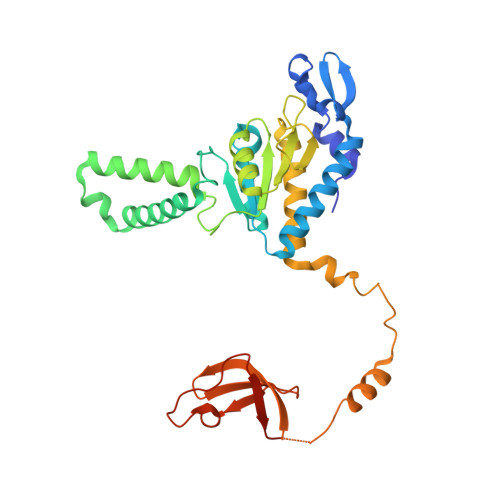
Structural basis for the interaction of protein S1 with the Escherichia coli ribosome.

Byrgazov, K.Grishkovskaya, I.Arenz, S.Coudevylle, N.Temmel, H.Wilson, D.N.Djinovic-Carugo, K.Moll, I.

(2015) Nucleic Acids Res 43: 661-673

  • DOI: https://doi.org/10.1093/nar/gku1314
  • Primary Citation of Related Structures:  
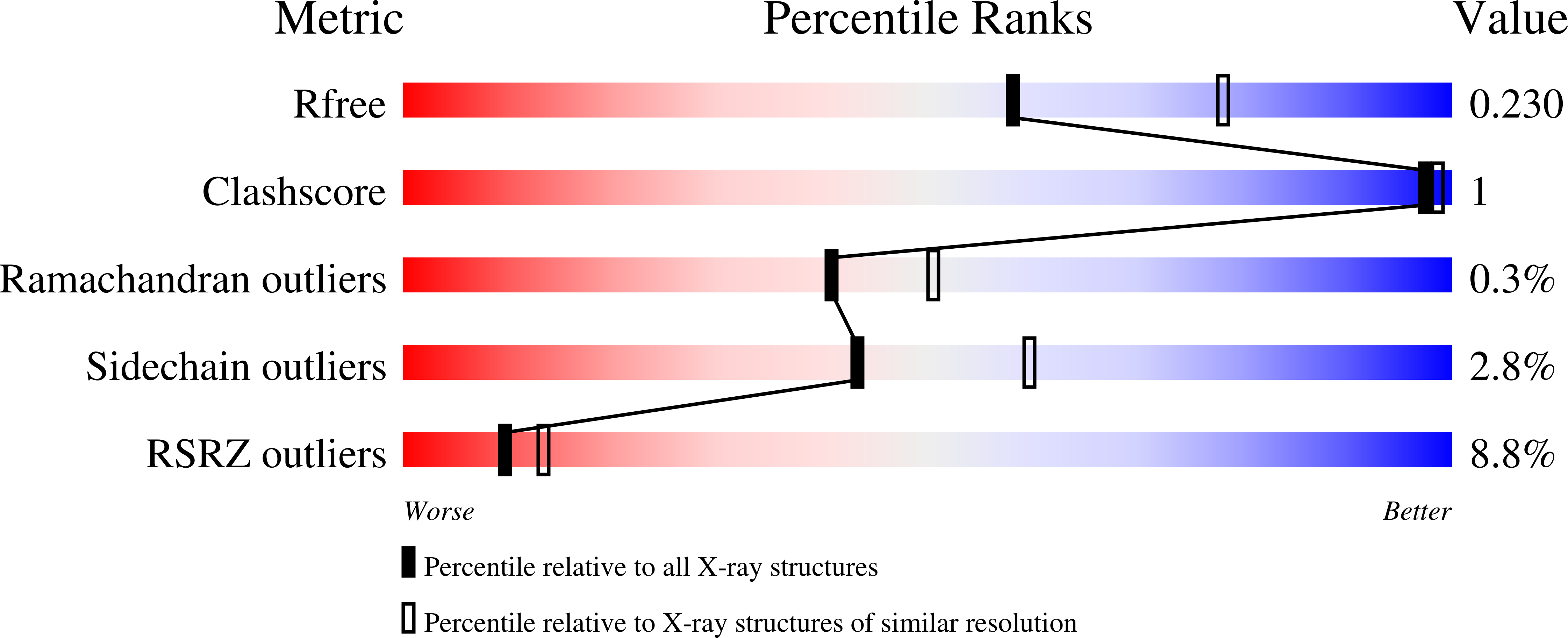
    4TOI

  • PubMed Abstract: 

    In Gram-negative bacteria, the multi-domain protein S1 is essential for translation initiation, as it recruits the mRNA and facilitates its localization in the decoding centre. In sharp contrast to its functional importance, S1 is still lacking from the high-resolution structures available for Escherichia coli and Thermus thermophilus ribosomes and thus the molecular mechanism governing the S1-ribosome interaction has still remained elusive. Here, we present the structure of the N-terminal S1 domain D1 when bound to the ribosome at atomic resolution by using a combination of NMR, X-ray crystallography and cryo-electron microscopy. Together with biochemical assays, the structure reveals that S1 is anchored to the ribosome primarily via a stabilizing π-stacking interaction within the short but conserved N-terminal segment that is flexibly connected to domain D1. This interaction is further stabilized by salt bridges involving the zinc binding pocket of protein S2. Overall, this work provides one hitherto enigmatic piece in the 'ribosome puzzle', namely the detailed molecular insight into the topology of the S1-ribosome interface. Moreover, our data suggest novel mechanisms that have the potential to modulate protein synthesis in response to environmental cues by changing the affinity of S1 for the ribosome.


  • Organizational Affiliation

    Department of Microbiology, Immunobiology and Genetics, Max F. Perutz Laboratories, Centre for Molecular Biology, University of Vienna, Dr. Bohrgasse 9/4, 1030 Vienna, Austria.


Macromolecules
Find similar proteins by:  (by identity cutoff)  |  3D Structure
Entity ID: 1
MoleculeChains Sequence LengthOrganismDetailsImage
30S ribosomal protein S2,Ribosomal protein S1326Escherichia coliEscherichia coli TA206
This entity is chimeric
Mutation(s): 0 
Gene Names: rpsBBU34_07500ECs0171LF82_1969ECKG_00790
UniProt
Find proteins for P0A7V0 (Escherichia coli (strain K12))
Explore P0A7V0 
Go to UniProtKB:  P0A7V0
Entity Groups  
Sequence Clusters30% Identity50% Identity70% Identity90% Identity95% Identity100% Identity
UniProt GroupP0A7V0
Sequence Annotations
Expand
  • Reference Sequence
Small Molecules
Ligands 1 Unique
IDChains Name / Formula / InChI Key2D Diagram3D Interactions
ZN
Query on ZN

Download Ideal Coordinates CCD File 
B [auth A]ZINC ION
Zn
PTFCDOFLOPIGGS-UHFFFAOYSA-N
Experimental Data & Validation

Experimental Data

  • Method: X-RAY DIFFRACTION
  • Resolution: 2.30 Å
  • R-Value Free: 0.229 
  • R-Value Work: 0.164 
  • R-Value Observed: 0.167 
  • Space Group: P 31 2 1
Unit Cell:
Length ( Å )Angle ( ˚ )
a = 87.28α = 90
b = 87.28β = 90
c = 94.36γ = 120
Software Package:
Software NamePurpose
XDSdata reduction
PDB_EXTRACTdata extraction
PHENIXrefinement
BALBESphasing
Aimlessdata scaling
XSCALEdata reduction

Structure Validation

View Full Validation Report


Entry History 

Deposition Data

Revision History  (Full details and data files)

  • Version 1.0: 2014-12-31
    Type: Initial release
  • Version 1.1: 2015-01-21
    Changes: Database references
  • Version 1.2: 2023-12-20
    Changes: Data collection, Database references, Refinement description