4TNW

C. elegans glutamate-gated chloride channel (GluCl) in complex with Fab and POPC in a lipid-modulated conformation


Experimental Data Snapshot

  • Method: X-RAY DIFFRACTION
  • Resolution: 3.20 Å
  • R-Value Free: 0.251 
  • R-Value Work: 0.227 
  • R-Value Observed: 0.228 

wwPDB Validation   3D Report Full Report


Ligand Structure Quality Assessment 


This is version 1.5 of the entry. See complete history


Literature

X-ray structures of GluCl in apo states reveal a gating mechanism of Cys-loop receptors.

Althoff, T.Hibbs, R.E.Banerjee, S.Gouaux, E.

(2014) Nature 512: 333-337

  • DOI: https://doi.org/10.1038/nature13669
  • Primary Citation of Related Structures:  
    4TNV, 4TNW

  • PubMed Abstract: 

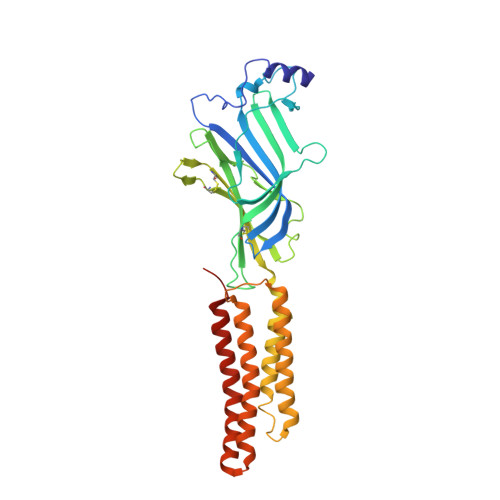
    Cys-loop receptors are neurotransmitter-gated ion channels that are essential mediators of fast chemical neurotransmission and are associated with a large number of neurological diseases and disorders, as well as parasitic infections. Members of this ion channel superfamily mediate excitatory or inhibitory neurotransmission depending on their ligand and ion selectivity. Structural information for Cys-loop receptors comes from several sources including electron microscopic studies of the nicotinic acetylcholine receptor, high-resolution X-ray structures of extracellular domains and X-ray structures of bacterial orthologues. In 2011 our group published structures of the Caenorhabditis elegans glutamate-gated chloride channel (GluCl) in complex with the allosteric partial agonist ivermectin, which provided insights into the structure of a possibly open state of a eukaryotic Cys-loop receptor, the basis for anion selectivity and channel block, and the mechanism by which ivermectin and related molecules stabilize the open state and potentiate neurotransmitter binding. However, there remain unanswered questions about the mechanism of channel opening and closing, the location and nature of the shut ion channel gate, the transitions between the closed/resting, open/activated and closed/desensitized states, and the mechanism by which conformational changes are coupled between the extracellular, orthosteric agonist binding domain and the transmembrane, ion channel domain. Here we present two conformationally distinct structures of C. elegans GluCl in the absence of ivermectin. Structural comparisons reveal a quaternary activation mechanism arising from rigid-body movements between the extracellular and transmembrane domains and a mechanism for modulation of the receptor by phospholipids.


  • Organizational Affiliation

    1] Vollum Institute, Oregon Health &Science University, 3181 SW Sam Jackson Park Road, Portland, Oregon 97239, USA [2] Department of Physiology, David Geffen School of Medicine, University of California Los Angeles, 10833 Le Conte Avenue, Los Angeles, California 90095-1751, USA (T.A.); Department of Neuroscience, University of Texas Southwestern Medical Center, 5323 Harry Hines Blvd, Dallas, Texas 75390-9111, USA (R.E.H.). [3].


Macromolecules
Find similar proteins by:  (by identity cutoff)  |  3D Structure
Entity ID: 1
MoleculeChains Sequence LengthOrganismDetailsImage
Avermectin-sensitive glutamate-gated chloride channel GluCl alpha347Caenorhabditis elegansMutation(s): 0 
Gene Names: glc-1CELE_F11A5.10F11A5.10
Membrane Entity: Yes 
UniProt
Find proteins for G5EBR3 (Caenorhabditis elegans)
Explore G5EBR3 
Go to UniProtKB:  G5EBR3
Entity Groups  
Sequence Clusters30% Identity50% Identity70% Identity90% Identity95% Identity100% Identity
UniProt GroupG5EBR3
Sequence Annotations
Expand
  • Reference Sequence
Find similar proteins by:  (by identity cutoff)  |  3D Structure
Entity ID: 2
MoleculeChains Sequence LengthOrganismDetailsImage
Mouse monoclonal Fab fragment, heavy chain224Mus musculusMutation(s): 0 
Entity Groups  
Sequence Clusters30% Identity50% Identity70% Identity90% Identity95% Identity100% Identity
Sequence Annotations
Expand
  • Reference Sequence
Find similar proteins by:  (by identity cutoff)  |  3D Structure
Entity ID: 3
MoleculeChains Sequence LengthOrganismDetailsImage
Mouse monoclonal Fab fragment, light chain215Mus musculusMutation(s): 0 
Entity Groups  
Sequence Clusters30% Identity50% Identity70% Identity90% Identity95% Identity100% Identity
Sequence Annotations
Expand
  • Reference Sequence
Small Molecules
Ligands 5 Unique
IDChains Name / Formula / InChI Key2D Diagram3D Interactions
POV
Query on POV

Download Ideal Coordinates CCD File 
BB [auth T]
FA [auth A]
MA [auth D]
TA [auth P]
VA [auth Q]
BB [auth T],
FA [auth A],
MA [auth D],
TA [auth P],
VA [auth Q],
XA [auth R],
YA [auth R]
(2S)-3-(hexadecanoyloxy)-2-[(9Z)-octadec-9-enoyloxy]propyl 2-(trimethylammonio)ethyl phosphate
C42 H82 N O8 P
WTJKGGKOPKCXLL-PFDVCBLKSA-N
LMT
Query on LMT

Download Ideal Coordinates CCD File 
AB [auth S]
DB [auth T]
JA [auth B]
LA [auth C]
OA [auth D]
AB [auth S],
DB [auth T],
JA [auth B],
LA [auth C],
OA [auth D],
QA [auth E],
SA [auth P]
DODECYL-BETA-D-MALTOSIDE
C24 H46 O11
NLEBIOOXCVAHBD-QKMCSOCLSA-N
NAG
Query on NAG

Download Ideal Coordinates CCD File 
CB [auth T]
EA [auth A]
IA [auth B]
KA [auth C]
NA [auth D]
CB [auth T],
EA [auth A],
IA [auth B],
KA [auth C],
NA [auth D],
PA [auth E],
RA [auth P],
WA [auth Q],
ZA [auth S]
2-acetamido-2-deoxy-beta-D-glucopyranose
C8 H15 N O6
OVRNDRQMDRJTHS-FMDGEEDCSA-N
GOL
Query on GOL

Download Ideal Coordinates CCD File 
HA [auth B]GLYCEROL
C3 H8 O3
PEDCQBHIVMGVHV-UHFFFAOYSA-N
CL
Query on CL

Download Ideal Coordinates CCD File 
GA [auth A],
UA [auth P]
CHLORIDE ION
Cl
VEXZGXHMUGYJMC-UHFFFAOYSA-M
Experimental Data & Validation

Experimental Data

  • Method: X-RAY DIFFRACTION
  • Resolution: 3.20 Å
  • R-Value Free: 0.251 
  • R-Value Work: 0.227 
  • R-Value Observed: 0.228 
  • Space Group: C 1 2 1
Unit Cell:
Length ( Å )Angle ( ˚ )
a = 453.75α = 90
b = 192.87β = 92.27
c = 196.14γ = 90
Software Package:
Software NamePurpose
PHENIXrefinement
ADSCdata collection
HKL-2000data reduction
HKL-2000data scaling
XDSdata reduction
XSCALEdata scaling
PHASERphasing
DMphasing
Cootmodel building

Structure Validation

View Full Validation Report


Ligand Structure Quality Assessment 


Entry History & Funding Information

Deposition Data


Funding OrganizationLocationGrant Number
National Institutes of Health/National Institute of General Medical Sciences (NIH/NIGMS)United States5 R01 GM100400
German Research Foundation (DFG)GermanyAL 1725-1/1
National Institutes of Health/National Institute of Neurological Disorders and Stroke (NIH/NINDS)United StatesF32NS061404

Revision History  (Full details and data files)

  • Version 1.0: 2014-08-13
    Type: Initial release
  • Version 1.1: 2014-09-03
    Changes: Database references
  • Version 1.2: 2017-09-27
    Changes: Author supporting evidence, Database references, Derived calculations, Other, Source and taxonomy, Structure summary
  • Version 1.3: 2019-12-18
    Changes: Author supporting evidence
  • Version 1.4: 2020-07-29
    Type: Remediation
    Reason: Carbohydrate remediation
    Changes: Data collection, Derived calculations, Refinement description, Structure summary
  • Version 1.5: 2023-09-27
    Changes: Data collection, Database references, Refinement description, Structure summary