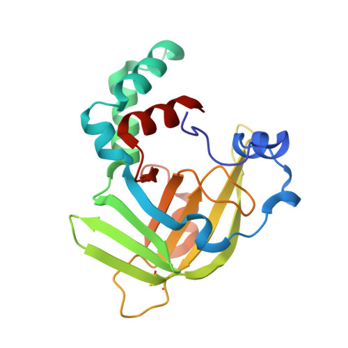
4TNN

Crystal structure of Escherichia coli protein YodA in complex with Ni - artifact of purification.


Experimental Data Snapshot

  • Method: X-RAY DIFFRACTION
  • Resolution: 1.95 Å
  • R-Value Free: 0.215 
  • R-Value Work: 0.167 
  • R-Value Observed: 0.169 

wwPDB Validation   3D Report Full Report


This is version 1.6 of the entry. See complete history


Literature

Protein purification and crystallization artifacts: The tale usually not told.

Niedzialkowska, E.Gasiorowska, O.Handing, K.B.Majorek, K.A.Porebski, P.J.Shabalin, I.G.Zasadzinska, E.Cymborowski, M.Minor, W.

(2016) Protein Sci 25: 720-733

  • DOI: https://doi.org/10.1002/pro.2861
  • Primary Citation of Related Structures:  
    4TNN, 4YYC, 4ZNZ

  • PubMed Abstract: 

    The misidentification of a protein sample, or contamination of a sample with the wrong protein, may be a potential reason for the non-reproducibility of experiments. This problem may occur in the process of heterologous overexpression and purification of recombinant proteins, as well as purification of proteins from natural sources. If the contaminated or misidentified sample is used for crystallization, in many cases the problem may not be detected until structures are determined. In the case of functional studies, the problem may not be detected for years. Here several procedures that can be successfully used for the identification of crystallized protein contaminants, including: (i) a lattice parameter search against known structures, (ii) sequence or fold identification from partially built models, and (iii) molecular replacement with common contaminants as search templates have been presented. A list of common contaminant structures to be used as alternative search models was provided. These methods were used to identify four cases of purification and crystallization artifacts. This report provides troubleshooting pointers for researchers facing difficulties in phasing or model building.


  • Organizational Affiliation

    Department of Molecular Physiology and Biological Physics, University of Virginia School of Medicine, 1340 Jefferson Park Avenue, Jordan Hall, Room 4223, Charlottesville, Virginia, 22908.


Macromolecules
Find similar proteins by:  (by identity cutoff)  |  3D Structure
Entity ID: 1
MoleculeChains Sequence LengthOrganismDetailsImage
Metal-binding lipocalin193Escherichia coli str. K-12 substr. MC4100Mutation(s): 0 
Gene Names: yodABN896_1774
Entity Groups  
Sequence Clusters30% Identity50% Identity70% Identity90% Identity95% Identity100% Identity
Sequence Annotations
Expand
  • Reference Sequence
Experimental Data & Validation

Experimental Data

Unit Cell:
Length ( Å )Angle ( ˚ )
a = 76.583α = 90
b = 76.583β = 90
c = 61.944γ = 120
Software Package:
Software NamePurpose
HKL-3000data collection
HKL-3000data reduction
HKL-3000data scaling
SHELXDEphasing
MLPHAREphasing
DMphasing
Cootmodel building
PDB_EXTRACTdata extraction
REFMACrefinement
DENZOdata reduction
SCALEPACKdata scaling

Structure Validation

View Full Validation Report


Entry History & Funding Information

Deposition Data


Funding OrganizationLocationGrant Number
National Institutes of Health/National Institute of General Medical Sciences (NIH/NIGMS)United StatesGM053163

Revision History  (Full details and data files)

  • Version 1.0: 2014-06-25
    Type: Initial release
  • Version 1.1: 2014-09-24
    Changes: Refinement description
  • Version 1.2: 2017-04-19
    Changes: Database references
  • Version 1.3: 2017-09-06
    Changes: Author supporting evidence, Refinement description
  • Version 1.4: 2019-12-25
    Changes: Author supporting evidence
  • Version 1.5: 2022-04-13
    Changes: Database references, Derived calculations, Structure summary
  • Version 1.6: 2023-09-27
    Changes: Data collection, Refinement description