4TLL

Crystal structure of GluN1/GluN2B NMDA receptor, structure 1


Experimental Data Snapshot

  • Method: X-RAY DIFFRACTION
  • Resolution: 3.59 Å
  • R-Value Free: 0.305 
  • R-Value Work: 0.266 
  • R-Value Observed: 0.268 

wwPDB Validation   3D Report Full Report


Ligand Structure Quality Assessment 


This is version 1.5 of the entry. See complete history


Literature

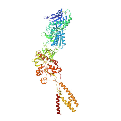
NMDA receptor structures reveal subunit arrangement and pore architecture.

Lee, C.H.Lu, W.Michel, J.C.Goehring, A.Du, J.Song, X.Gouaux, E.

(2014) Nature 511: 191-197

  • DOI: https://doi.org/10.1038/nature13548
  • Primary Citation of Related Structures:  
    4TLL, 4TLM

  • PubMed Abstract: 

    N-methyl-d-aspartate (NMDA) receptors are Hebbian-like coincidence detectors, requiring binding of glycine and glutamate in combination with the relief of voltage-dependent magnesium block to open an ion conductive pore across the membrane bilayer. Despite the importance of the NMDA receptor in the development and function of the brain, a molecular structure of an intact receptor has remained elusive. Here we present X-ray crystal structures of the Xenopus laevis GluN1-GluN2B NMDA receptor with the allosteric inhibitor, Ro25-6981, partial agonists and the ion channel blocker, MK-801. Receptor subunits are arranged in a 1-2-1-2 fashion, demonstrating extensive interactions between the amino-terminal and ligand-binding domains. The transmembrane domains harbour a closed-blocked ion channel, a pyramidal central vestibule lined by residues implicated in binding ion channel blockers and magnesium, and a ∼twofold symmetric arrangement of ion channel pore loops. These structures provide new insights into the architecture, allosteric coupling and ion channel function of NMDA receptors.


  • Organizational Affiliation

    1] Vollum Institute, Oregon Health & Science University, 3181 SW Sam Jackson Park Road, Portland, Oregon 97239, USA [2].


Macromolecules
Find similar proteins by:  (by identity cutoff)  |  3D Structure
Entity ID: 1
MoleculeChains Sequence LengthOrganismDetailsImage
receptor subunit GluN1
A, C
823Xenopus laevisMutation(s): 15 
Membrane Entity: Yes 
UniProt
Find proteins for A0A1L8F5J9 (Xenopus laevis)
Explore A0A1L8F5J9 
Go to UniProtKB:  A0A1L8F5J9
Entity Groups  
Sequence Clusters30% Identity50% Identity70% Identity90% Identity95% Identity100% Identity
UniProt GroupA0A1L8F5J9
Sequence Annotations
Expand
  • Reference Sequence
Find similar proteins by:  (by identity cutoff)  |  3D Structure
Entity ID: 2
MoleculeChains Sequence LengthOrganismDetailsImage
receptor subunit GluN2B
B, D
824Xenopus laevisMutation(s): 11 
Gene Names: NR2B
Membrane Entity: Yes 
UniProt
Find proteins for A7XY94 (Xenopus laevis)
Explore A7XY94 
Go to UniProtKB:  A7XY94
Entity Groups  
Sequence Clusters30% Identity50% Identity70% Identity90% Identity95% Identity100% Identity
UniProt GroupA7XY94
Sequence Annotations
Expand
  • Reference Sequence
Small Molecules
Ligands 4 Unique
IDChains Name / Formula / InChI Key2D Diagram3D Interactions
QEM
Query on QEM

Download Ideal Coordinates CCD File 
I [auth B],
K [auth D]
4-[(1R,2S)-3-(4-benzylpiperidin-1-yl)-1-hydroxy-2-methylpropyl]phenol
C22 H29 N O2
WVZSEUPGUDIELE-HTAPYJJXSA-N
NAG
Query on NAG

Download Ideal Coordinates CCD File 
E [auth A],
F [auth A],
G [auth A],
J [auth B]
2-acetamido-2-deoxy-beta-D-glucopyranose
C8 H15 N O6
OVRNDRQMDRJTHS-FMDGEEDCSA-N
JEG
Query on JEG

Download Ideal Coordinates CCD File 
L [auth D]trans-1-aminocyclobutane-1,3-dicarboxylic acid
C6 H9 N O4
GGMYWPBNZXRMME-HSRNZHMGSA-N
1AC
Query on 1AC

Download Ideal Coordinates CCD File 
H [auth A]1-AMINOCYCLOPROPANECARBOXYLIC ACID
C4 H7 N O2
PAJPWUMXBYXFCZ-UHFFFAOYSA-N
Binding Affinity Annotations 
IDSourceBinding Affinity
QEM BindingDB:  4TLL Ki: min: 6, max: 173 (nM) from 3 assay(s)
IC50: min: 6, max: 5.20e+4 (nM) from 6 assay(s)
Experimental Data & Validation

Experimental Data

  • Method: X-RAY DIFFRACTION
  • Resolution: 3.59 Å
  • R-Value Free: 0.305 
  • R-Value Work: 0.266 
  • R-Value Observed: 0.268 
  • Space Group: C 1 2 1
Unit Cell:
Length ( Å )Angle ( ˚ )
a = 201.46α = 90
b = 117.26β = 106.7
c = 218.76γ = 90
Software Package:
Software NamePurpose
PHENIXrefinement

Structure Validation

View Full Validation Report


Ligand Structure Quality Assessment 


Entry History 

Deposition Data

Revision History  (Full details and data files)

  • Version 1.0: 2014-07-02
    Type: Initial release
  • Version 1.1: 2014-07-09
    Changes: Database references, Structure summary
  • Version 1.2: 2014-07-16
    Changes: Database references
  • Version 1.3: 2014-09-24
    Changes: Database references
  • Version 1.4: 2020-07-29
    Type: Remediation
    Reason: Carbohydrate remediation
    Changes: Data collection, Database references, Derived calculations, Other, Refinement description, Source and taxonomy, Structure summary
  • Version 1.5: 2023-12-27
    Changes: Data collection, Database references, Structure summary