4THN

THE CRYSTAL STRUCTURE OF ALPHA-THROMBIN-HIRUNORM IV COMPLEX REVEALS A NOVEL SPECIFICITY SITE RECOGNITION MODE.


Experimental Data Snapshot

  • Method: X-RAY DIFFRACTION
  • Resolution: 2.50 Å
  • R-Value Work: 0.173 

wwPDB Validation   3D Report Full Report


This is version 1.4 of the entry. See complete history


Literature

The crystal structure of alpha-thrombin-hirunorm IV complex reveals a novel specificity site recognition mode.

Lombardi, A.De Simone, G.Nastri, F.Galdiero, S.Della Morte, R.Staiano, N.Pedone, C.Bolognesi, M.Pavone, V.

(1999) Protein Sci 8: 91-95

  • DOI: https://doi.org/10.1110/ps.8.1.91
  • Primary Citation of Related Structures:  
    4THN

  • PubMed Abstract: 

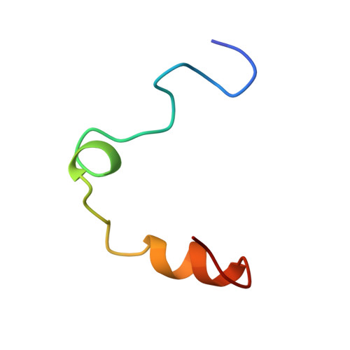
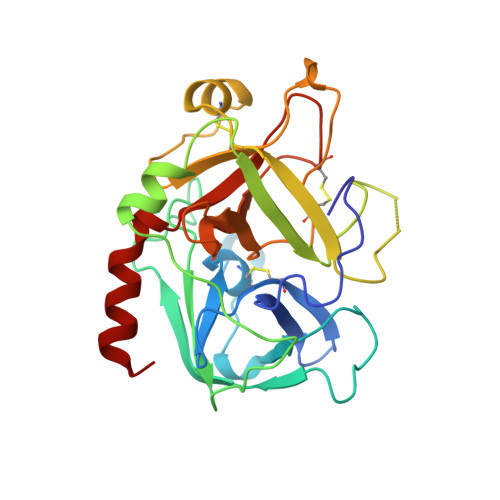
    The X-ray crystal structure of the human alpha-thrombin-hirunorm IV complex has been determined at 2.5 A resolution, and refined to an R-factor of 0.173. The structure reveals an inhibitor binding mode distinctive of a true hirudin mimetic, which justifies the high inhibitory potency and the selectivity of hirunorm IV. This novel inhibitor, composed of 26 amino acids, interacts through the N-terminal end with the alpha-thrombin active site in a nonsubstrate mode, and binds specifically to the fibrinogen recognition exosite through the C-terminal end. The backbone of the N-terminal tripeptide Chg1"-Arg2"-2Na13" (Chg, cyclohexyl-glycine; 2Na1, beta-(2-naphthyl)-alanine) forms a parallel beta-strand to the thrombin main-chain segment Ser214-Gly216. The Chg1" side chain occupies the S2 site, Arg2" penetrates into the S1 specificity site, while the 2Na13" side chain occupies the aryl binding site. The Arg2" side chain enters the S1 specificity pocket from a position quite apart from the canonical P1 site. This notwithstanding, the Arg2" side chain establishes the typical ion pair with the carboxylate group of Asp189.


  • Organizational Affiliation

    Centro Interuniversitario di Ricerca su Peptidi Bioattivi & Centro di Studio di Biocristallografia-CNR, University of Napoli Federico II, Italy.


Macromolecules
Find similar proteins by:  (by identity cutoff)  |  3D Structure
Entity ID: 1
MoleculeChains Sequence LengthOrganismDetailsImage
ALPHA-THROMBINA [auth L]36Homo sapiensMutation(s): 0 
EC: 3.4.21.5
UniProt & NIH Common Fund Data Resources
Find proteins for P00734 (Homo sapiens)
Explore P00734 
Go to UniProtKB:  P00734
PHAROS:  P00734
GTEx:  ENSG00000180210 
Entity Groups  
Sequence Clusters30% Identity50% Identity70% Identity90% Identity95% Identity100% Identity
UniProt GroupP00734
Sequence Annotations
Expand
  • Reference Sequence
Find similar proteins by:  (by identity cutoff)  |  3D Structure
Entity ID: 2
MoleculeChains Sequence LengthOrganismDetailsImage
ALPHA-THROMBINB [auth H]259Homo sapiensMutation(s): 0 
EC: 3.4.21.5
UniProt & NIH Common Fund Data Resources
Find proteins for P00734 (Homo sapiens)
Explore P00734 
Go to UniProtKB:  P00734
PHAROS:  P00734
GTEx:  ENSG00000180210 
Entity Groups  
Sequence Clusters30% Identity50% Identity70% Identity90% Identity95% Identity100% Identity
UniProt GroupP00734
Sequence Annotations
Expand
  • Reference Sequence
Find similar proteins by:  (by identity cutoff)  |  3D Structure
Entity ID: 3
MoleculeChains Sequence LengthOrganismDetailsImage
HIRUNORM IVC [auth I]26N/AMutation(s): 0 
Entity Groups  
Sequence Clusters30% Identity50% Identity70% Identity90% Identity95% Identity100% Identity
Sequence Annotations
Expand
  • Reference Sequence
Small Molecules
Ligands 1 Unique
IDChains Name / Formula / InChI Key2D Diagram3D Interactions
NAG
Query on NAG

Download Ideal Coordinates CCD File 
D [auth H]2-acetamido-2-deoxy-beta-D-glucopyranose
C8 H15 N O6
OVRNDRQMDRJTHS-FMDGEEDCSA-N
Modified Residues  3 Unique
IDChains TypeFormula2D DiagramParent
AIB
Query on AIB
C [auth I]L-PEPTIDE LINKINGC4 H9 N O2ALA
ALC
Query on ALC
C [auth I]L-PEPTIDE LINKINGC9 H17 N O2ALA
NAL
Query on NAL
C [auth I]L-PEPTIDE LINKINGC13 H13 N O2ALA
Experimental Data & Validation

Experimental Data

  • Method: X-RAY DIFFRACTION
  • Resolution: 2.50 Å
  • R-Value Work: 0.173 
  • Space Group: C 1 2 1
Unit Cell:
Length ( Å )Angle ( ˚ )
a = 72.3α = 90
b = 72.7β = 100
c = 73.6γ = 90
Software Package:
Software NamePurpose
MOSFLMdata reduction
CCP4data reduction
CCP4model building
TNTrefinement
CCP4data scaling
CCP4phasing

Structure Validation

View Full Validation Report


Entry History 

Deposition Data

Revision History  (Full details and data files)

  • Version 1.0: 1999-06-15
    Type: Initial release
  • Version 1.1: 2008-03-25
    Changes: Version format compliance
  • Version 1.2: 2011-07-13
    Changes: Atomic model, Database references, Derived calculations, Non-polymer description, Structure summary, Version format compliance
  • Version 1.3: 2020-07-29
    Type: Remediation
    Reason: Carbohydrate remediation
    Changes: Advisory, Data collection, Derived calculations, Structure summary
  • Version 1.4: 2023-08-09
    Changes: Advisory, Database references, Refinement description, Structure summary