


Multiple mechanisms render Erk proteins MEK-independent
Livnah, O., Karamansha, Y.To be published.
Experimental Data Snapshot
wwPDB Validation 3D Report Full Report

Entity ID: 1 | |||||
|---|---|---|---|---|---|
| Molecule | Chains | Sequence Length | Organism | Details | Image |
| Mitogen-activated protein kinase 1 | 358 | Rattus norvegicus | Mutation(s): 0 Gene Names: Mapk1, Erk2, Mapk, Prkm1 EC: 2.7.11.24 | 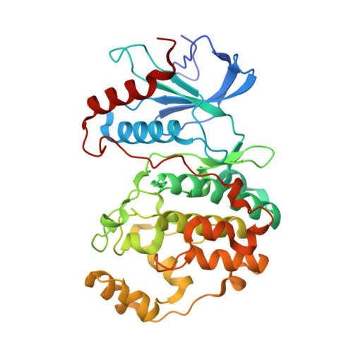 | |
UniProt | |||||
Find proteins for P63086 (Rattus norvegicus) Explore P63086 Go to UniProtKB: P63086 | |||||
Entity Groups | |||||
| Sequence Clusters | 30% Identity50% Identity70% Identity90% Identity95% Identity100% Identity | ||||
| UniProt Group | P63086 | ||||
Sequence AnnotationsExpand | |||||
| |||||
| Length ( Å ) | Angle ( ˚ ) |
|---|---|
| a = 48.846 | α = 90 |
| b = 70.242 | β = 109.66 |
| c = 60.744 | γ = 90 |
| Software Name | Purpose |
|---|---|
| HKL-2000 | data collection |
| AMoRE | phasing |
| REFMAC | refinement |
| HKL-2000 | data reduction |
| SCALEPACK | data scaling |
RCSB PDB (citation) is hosted by
RCSB PDB is a member of the

