


Crystal Structure of Phosphoglycerate Oxidoreductase from Vibrio Cholerae O395
Tarique, K.F., Rehman, S.A.A., Devi, S., Gourinath, S.To be published.
Experimental Data Snapshot
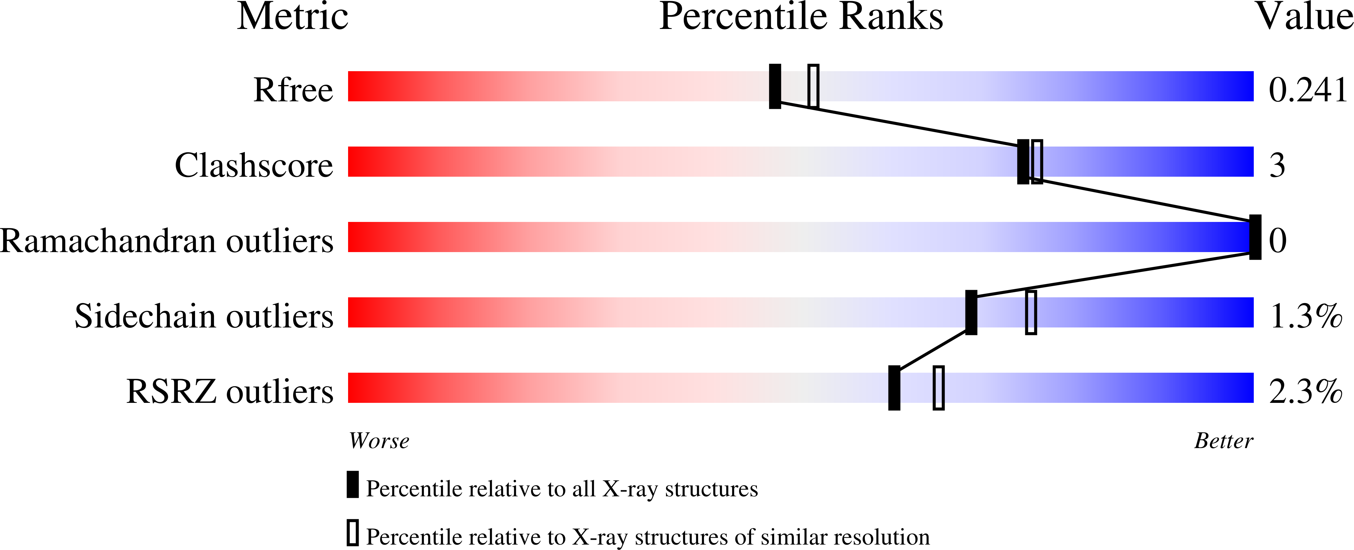
wwPDB Validation 3D Report Full Report
Entity ID: 1 | |||||
|---|---|---|---|---|---|
| Molecule | Chains | Sequence Length | Organism | Details | Image |
| D-3-phosphoglycerate dehydrogenase-related protein | A [auth D], B [auth A], C [auth B], D [auth C] | 332 | Vibrio cholerae O395 | Mutation(s): 0 Gene Names: VC0395_0573, VC395_A0682 EC: 1.1.1.95 | 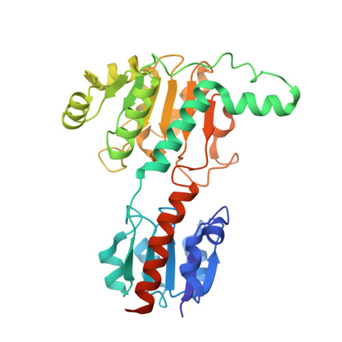 |
UniProt | |||||
Find proteins for A0A0H2UKZ7 (Vibrio cholerae serotype O1 (strain ATCC 39541 / Classical Ogawa 395 / O395)) Explore A0A0H2UKZ7 Go to UniProtKB: A0A0H2UKZ7 | |||||
Entity Groups | |||||
| Sequence Clusters | 30% Identity50% Identity70% Identity90% Identity95% Identity100% Identity | ||||
| UniProt Group | A0A0H2UKZ7 | ||||
Sequence AnnotationsExpand | |||||
| |||||
| Length ( Å ) | Angle ( ˚ ) |
|---|---|
| a = 51.916 | α = 64.39 |
| b = 83.258 | β = 81.94 |
| c = 85.986 | γ = 76.03 |
| Software Name | Purpose |
|---|---|
| MAR345dtb | data collection |
| PHASER | phasing |
| REFMAC | refinement |
| HKL-2000 | data reduction |
| HKL-2000 | data scaling |
RCSB PDB (citation) is hosted by
RCSB PDB is a member of the

