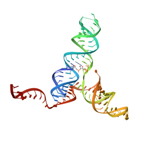
Structural analysis of a class III preQ1 riboswitch reveals an aptamer distant from a ribosome-binding site regulated by fast dynamics.
Liberman, J.A., Suddala, K.C., Aytenfisu, A., Chan, D., Belashov, I.A., Salim, M., Mathews, D.H., Spitale, R.C., Walter, N.G., Wedekind, J.E.(2015) Proc Natl Acad Sci U S A 112: E3485-E3494
- PubMed: 26106162
- DOI: https://doi.org/10.1073/pnas.1503955112
- Primary Citation of Related Structures:
4RZD - PubMed Abstract:
PreQ1-III riboswitches are newly identified RNA elements that control bacterial genes in response to preQ1 (7-aminomethyl-7-deazaguanine), a precursor to the essential hypermodified tRNA base queuosine. Although numerous riboswitches fold as H-type or HLout-type pseudoknots that integrate ligand-binding and regulatory sequences within a single folded domain, the preQ1-III riboswitch aptamer forms a HLout-type pseudoknot that does not appear to incorporate its ribosome-binding site (RBS). To understand how this unusual organization confers function, we determined the crystal structure of the class III preQ1 riboswitch from Faecalibacterium prausnitzii at 2.75 Å resolution. PreQ1 binds tightly (KD,app 6.5 ± 0.5 nM) between helices P1 and P2 of a three-way helical junction wherein the third helix, P4, projects orthogonally from the ligand-binding pocket, exposing its stem-loop to base pair with the 3' RBS. Biochemical analysis, computational modeling, and single-molecule FRET imaging demonstrated that preQ1 enhances P4 reorientation toward P1-P2, promoting a partially nested, H-type pseudoknot in which the RBS undergoes rapid docking (kdock ∼ 0.6 s(-1)) and undocking (kundock ∼ 1.1 s(-1)). Discovery of such dynamic conformational switching provides insight into how a riboswitch with bipartite architecture uses dynamics to modulate expression platform accessibility, thus expanding the known repertoire of gene control strategies used by regulatory RNAs.
Organizational Affiliation:
Department of Biochemistry and Biophysics, and Center for RNA Biology, University of Rochester School of Medicine and Dentistry, Rochester, NY 14642;