Roles of intramolecular and intermolecular interactions in functional regulation of the Hsp70 J-protein co-chaperone Sis1.
Yu, H.Y., Ziegelhoffer, T., Osipiuk, J., Ciesielski, S.J., Baranowski, M., Zhou, M., Joachimiak, A., Craig, E.A.(2015) J Mol Biol 427: 1632-1643
- PubMed: 25687964
- DOI: https://doi.org/10.1016/j.jmb.2015.02.007
- Primary Citation of Related Structures:
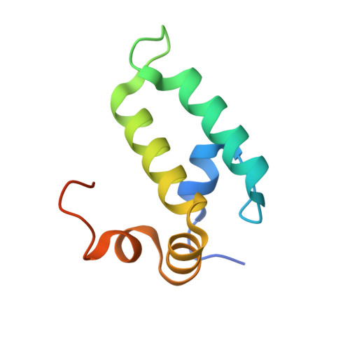
4RWU - PubMed Abstract:
Unlike other Hsp70 molecular chaperones, those of the eukaryotic cytosol have four residues, EEVD, at their C-termini. EEVD(Hsp70) binds adaptor proteins of the Hsp90 chaperone system and mitochondrial membrane preprotein receptors, thereby facilitating processing of Hsp70-bound clients through protein folding and translocation pathways. Among J-protein co-chaperones functioning in these pathways, Sis1 is unique, as it also binds the EEVD(Hsp70) motif. However, little is known about the role of the Sis1:EEVD(Hsp70) interaction. We found that deletion of EEVD(Hsp70) abolished the ability of Sis1, but not the ubiquitous J-protein Ydj1, to partner with Hsp70 in in vitro protein refolding. Sis1 co-chaperone activity with Hsp70∆EEVD was restored upon substitution of a glutamic acid of the J-domain. Structural analysis revealed that this key glutamic acid, which is not present in Ydj1, forms a salt bridge with an arginine of the immediately adjacent glycine-rich region. Thus, restoration of Sis1 in vitro activity suggests that intramolecular interactions between the J-domain and glycine-rich region control co-chaperone activity, which is optimal only when Sis1 interacts with the EEVD(Hsp70) motif. However, we found that disruption of the Sis1:EEVD(Hsp70) interaction enhances the ability of Sis1 to substitute for Ydj1 in vivo. Our results are consistent with the idea that interaction of Sis1 with EEVD(Hsp70) minimizes transfer of Sis1-bound clients to Hsp70s that are primed for client transfer to folding and translocation pathways by their preassociation with EEVD binding adaptor proteins. These interactions may be one means by which cells triage Ydj1- and Sis1-bound clients to productive and quality control pathways, respectively.
Organizational Affiliation:
Department of Biochemistry, University of Wisconsin-Madison, 433 Babcock Drive, Madison, WI 53706, USA.