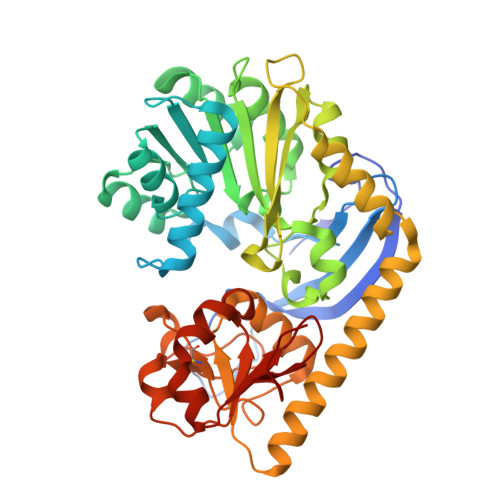
Structural Insight into MtmC, a Bifunctional Ketoreductase-Methyltransferase Involved in the Assembly of the Mithramycin Trisaccharide Chain.
Chen, J.M., Hou, C., Wang, G., Tsodikov, O.V., Rohr, J.(2015) Biochemistry 54: 2481-2489
- PubMed: 25587924
- DOI: https://doi.org/10.1021/bi501462g
- Primary Citation of Related Structures:
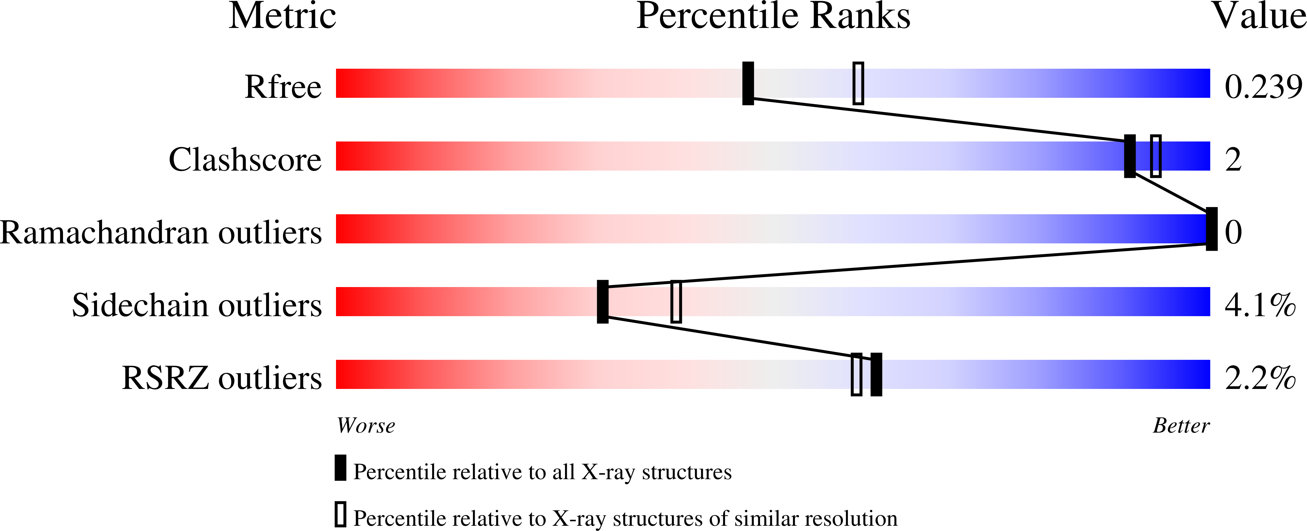
4RV9, 4RVD, 4RVF, 4RVG, 4RVH - PubMed Abstract:
More and more post-PKS tailoring enzymes are recognized as being multifunctional and codependent on other tailoring enzymes. One of the recently discovered intriguing examples is MtmC, a bifunctional TDP-4-keto-d-olivose ketoreductase-methyltransferase, which-in codependence with glycosyltransferase MtmGIV-is a key contributor to the biosynthesis of the critical trisaccharide chain of the antitumor antibiotic mithramycin (MTM), produced by Streptomyces argillaceus. We report crystal structures of three binary complexes of MtmC with its methylation cosubstrate SAM, its coproduct SAH, and a nucleotide TDP as well as crystal structures of two ternary complexes, MtmC-SAH-TDP-4-keto-d-olivose and MtmC-SAM-TDP, in the range of 2.2-2.7 Å resolution. The structures reveal general and sugar-specific recognition and catalytic structural features of MtmC. Depending on the catalytic function that is conducted by MtmC, it must bind either NADPH or SAM in the same cofactor binding pocket. A tyrosine residue (Tyr79) appears as a lid covering the sugar moiety of the substrate during the methyl transfer reaction. This residue swings out of the active site by ~180° in the absence of the substrate. This unique conformational change likely serves to release the methylated product and, possibly, to open the active site for binding the bulkier cosubstrate NADPH prior to the reduction reaction.
Organizational Affiliation:
Department of Pharmaceutical Sciences, College of Pharmacy, University of Kentucky, 789 South Limestone Street, Lexington, Kentucky 40536-0596, United States.