4RT0

Structure of the Alg44 PilZ domain from Pseudomonas aeruginosa PAO1 in complex with c-di-GMP


Experimental Data Snapshot

  • Method: X-RAY DIFFRACTION
  • Resolution: 1.80 Å
  • R-Value Free: 0.222 
  • R-Value Work: 0.187 
  • R-Value Observed: 0.189 

wwPDB Validation   3D Report Full Report


Ligand Structure Quality Assessment 


This is version 1.1 of the entry. See complete history


Literature

Dimeric c-di-GMP Is Required for Post-translational Regulation of Alginate Production in Pseudomonas aeruginosa.

Whitney, J.C.Whitfield, G.B.Marmont, L.S.Yip, P.Neculai, A.M.Lobsanov, Y.D.Robinson, H.Ohman, D.E.Howell, P.L.

(2015) J Biol Chem 290: 12451-12462

  • DOI: https://doi.org/10.1074/jbc.M115.645051
  • Primary Citation of Related Structures:  
    4RT0, 4RT1

  • PubMed Abstract: 

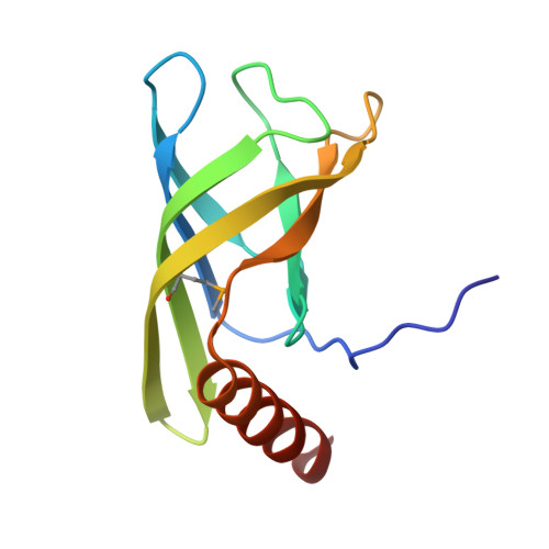
    Pseudomonas aeruginosa is an opportunistic human pathogen that secretes the exopolysaccharide alginate during infection of the respiratory tract of individuals afflicted with cystic fibrosis and chronic obstructive pulmonary disease. Among the proteins required for alginate production, Alg44 has been identified as an inner membrane protein whose bis-(3',5')-cyclic dimeric guanosine monophosphate (c-di-GMP) binding activity post-translationally regulates alginate secretion. In this study, we report the 1.8 Å crystal structure of the cytoplasmic region of Alg44 in complex with dimeric self-intercalated c-di-GMP and characterize its dinucleotide-binding site using mutational analysis. The structure shows that the c-di-GMP binding region of Alg44 adopts a PilZ domain fold with a dimerization mode not previously observed for this family of proteins. Calorimetric binding analysis of residues in the c-di-GMP binding site demonstrate that mutation of Arg-17 and Arg-95 alters the binding stoichiometry between c-di-GMP and Alg44 from 2:1 to 1:1. Introduction of these mutant alleles on the P. aeruginosa chromosome show that the residues required for binding of dimeric c-di-GMP in vitro are also required for efficient alginate production in vivo. These results suggest that the dimeric form of c-di-GMP represents the biologically active signaling molecule needed for the secretion of an important virulence factor produced by P. aeruginosa.


  • Organizational Affiliation

    From the Program in Molecular Structure and Function, Hospital for Sick Children, Toronto, Ontario M5G 0A4, Canada, the Department of Biochemistry, University of Toronto, Toronto, Ontario M5S 1A8, Canada.


Macromolecules
Find similar proteins by:  (by identity cutoff)  |  3D Structure
Entity ID: 1
MoleculeChains Sequence LengthOrganismDetailsImage
Alginate biosynthesis protein Alg44
A, B, C
112Pseudomonas aeruginosa PAO1Mutation(s): 1 
Gene Names: alg44PA3542
UniProt
Find proteins for Q9HY69 (Pseudomonas aeruginosa (strain ATCC 15692 / DSM 22644 / CIP 104116 / JCM 14847 / LMG 12228 / 1C / PRS 101 / PAO1))
Explore Q9HY69 
Go to UniProtKB:  Q9HY69
Entity Groups  
Sequence Clusters30% Identity50% Identity70% Identity90% Identity95% Identity100% Identity
UniProt GroupQ9HY69
Sequence Annotations
Expand
  • Reference Sequence
Small Molecules
Ligands 2 Unique
IDChains Name / Formula / InChI Key2D Diagram3D Interactions
C2E
Query on C2E

Download Ideal Coordinates CCD File 
D [auth A]
E [auth A]
F [auth B]
G [auth B]
H [auth C]
D [auth A],
E [auth A],
F [auth B],
G [auth B],
H [auth C],
I [auth C]
9,9'-[(2R,3R,3aS,5S,7aR,9R,10R,10aS,12S,14aR)-3,5,10,12-tetrahydroxy-5,12-dioxidooctahydro-2H,7H-difuro[3,2-d:3',2'-j][1,3,7,9,2,8]tetraoxadiphosphacyclododecine-2,9-diyl]bis(2-amino-1,9-dihydro-6H-purin-6-one)
C20 H24 N10 O14 P2
PKFDLKSEZWEFGL-MHARETSRSA-N
EDO
Query on EDO

Download Ideal Coordinates CCD File 
J [auth C],
K [auth C]
1,2-ETHANEDIOL
C2 H6 O2
LYCAIKOWRPUZTN-UHFFFAOYSA-N
Modified Residues  1 Unique
IDChains TypeFormula2D DiagramParent
MSE
Query on MSE
A, B, C
L-PEPTIDE LINKINGC5 H11 N O2 SeMET
Binding Affinity Annotations 
IDSourceBinding Affinity
C2E Binding MOAD:  4RT0 Kd: 3130 (nM) from 1 assay(s)
Experimental Data & Validation

Experimental Data

  • Method: X-RAY DIFFRACTION
  • Resolution: 1.80 Å
  • R-Value Free: 0.222 
  • R-Value Work: 0.187 
  • R-Value Observed: 0.189 
  • Space Group: P 31 2 1
Unit Cell:
Length ( Å )Angle ( ˚ )
a = 57.253α = 90
b = 57.253β = 90
c = 178.35γ = 120
Software Package:
Software NamePurpose
HKL-2000data collection
SOLVEphasing
RESOLVEmodel building
PHENIXrefinement
HKL-2000data reduction
HKL-2000data scaling
RESOLVEphasing

Structure Validation

View Full Validation Report


Ligand Structure Quality Assessment 


Entry History 

Deposition Data

Revision History  (Full details and data files)

  • Version 1.0: 2015-04-08
    Type: Initial release
  • Version 1.1: 2015-06-03
    Changes: Database references