4RR2

Crystal structure of human primase


Experimental Data Snapshot

  • Method: X-RAY DIFFRACTION
  • Resolution: 2.65 Å
  • R-Value Free: 0.271 
  • R-Value Work: 0.229 
  • R-Value Observed: 0.229 

wwPDB Validation   3D Report Full Report


This is version 1.2 of the entry. See complete history


Literature

Crystal structure of the human primase.

Baranovskiy, A.G.Zhang, Y.Suwa, Y.Babayeva, N.D.Gu, J.Pavlov, Y.I.Tahirov, T.H.

(2015) J Biol Chem 290: 5635-5646

  • DOI: https://doi.org/10.1074/jbc.M114.624742
  • Primary Citation of Related Structures:  
    4RR2

  • PubMed Abstract: 

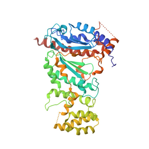
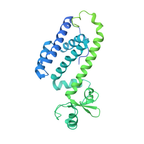
    DNA replication in bacteria and eukaryotes requires the activity of DNA primase, a DNA-dependent RNA polymerase that lays short RNA primers for DNA polymerases. Eukaryotic and archaeal primases are heterodimers consisting of small catalytic and large accessory subunits, both of which are necessary for RNA primer synthesis. Understanding of RNA synthesis priming in eukaryotes is currently limited due to the lack of crystal structures of the full-length primase and its complexes with substrates in initiation and elongation states. Here we report the crystal structure of the full-length human primase, revealing the precise overall organization of the enzyme, the relative positions of its functional domains, and the mode of its interaction with modeled DNA and RNA. The structure indicates that the dramatic conformational changes in primase are necessary to accomplish the initiation and then elongation of RNA synthesis. The presence of a long linker between the N- and C-terminal domains of p58 provides the structural basis for the bulk of enzyme's conformational flexibility. Deletion of most of this linker affected the initiation and elongation steps of the primer synthesis.


  • Organizational Affiliation

    From the Eppley Institute for Research in Cancer and Allied Diseases, University of Nebraska Medical Center, Omaha, Nebraska 68198.


Macromolecules
Find similar proteins by:  (by identity cutoff)  |  3D Structure
Entity ID: 1
MoleculeChains Sequence LengthOrganismDetailsImage
DNA primase small subunit
A, C
420Homo sapiensMutation(s): 0 
Gene Names: PRIM1
EC: 2.7.7
UniProt & NIH Common Fund Data Resources
Find proteins for P49642 (Homo sapiens)
Explore P49642 
Go to UniProtKB:  P49642
PHAROS:  P49642
GTEx:  ENSG00000198056 
Entity Groups  
Sequence Clusters30% Identity50% Identity70% Identity90% Identity95% Identity100% Identity
UniProt GroupP49642
Sequence Annotations
Expand
  • Reference Sequence
Find similar proteins by:  (by identity cutoff)  |  3D Structure
Entity ID: 2
MoleculeChains Sequence LengthOrganismDetailsImage
DNA primase large subunit
B, D
509Homo sapiensMutation(s): 0 
Gene Names: PRIM2PRIM2A
EC: 2.7.7
UniProt & NIH Common Fund Data Resources
Find proteins for P49643 (Homo sapiens)
Explore P49643 
Go to UniProtKB:  P49643
PHAROS:  P49643
GTEx:  ENSG00000146143 
Entity Groups  
Sequence Clusters30% Identity50% Identity70% Identity90% Identity95% Identity100% Identity
UniProt GroupP49643
Sequence Annotations
Expand
  • Reference Sequence
Experimental Data & Validation

Experimental Data

  • Method: X-RAY DIFFRACTION
  • Resolution: 2.65 Å
  • R-Value Free: 0.271 
  • R-Value Work: 0.229 
  • R-Value Observed: 0.229 
  • Space Group: P 1
Unit Cell:
Length ( Å )Angle ( ˚ )
a = 86.195α = 93.82
b = 88.899β = 96.57
c = 94.682γ = 111.72
Software Package:
Software NamePurpose
PHENIXmodel building
CNSrefinement
HKL-2000data reduction
HKL-2000data scaling
PHENIXphasing

Structure Validation

View Full Validation Report


Entry History 

Deposition Data

Revision History  (Full details and data files)

  • Version 1.0: 2015-01-21
    Type: Initial release
  • Version 1.1: 2015-03-11
    Changes: Database references
  • Version 1.2: 2024-02-28
    Changes: Data collection, Database references, Derived calculations