Structure of l-Serine Dehydratase from Legionella pneumophila: Novel Use of the C-Terminal Cysteine as an Intrinsic Competitive Inhibitor.
Thoden, J.B., Holden, H.M., Grant, G.A.(2014) Biochemistry 53: 7615-7624
- PubMed: 25380533
- DOI: https://doi.org/10.1021/bi501253w
- Primary Citation of Related Structures:
4RQO - PubMed Abstract:
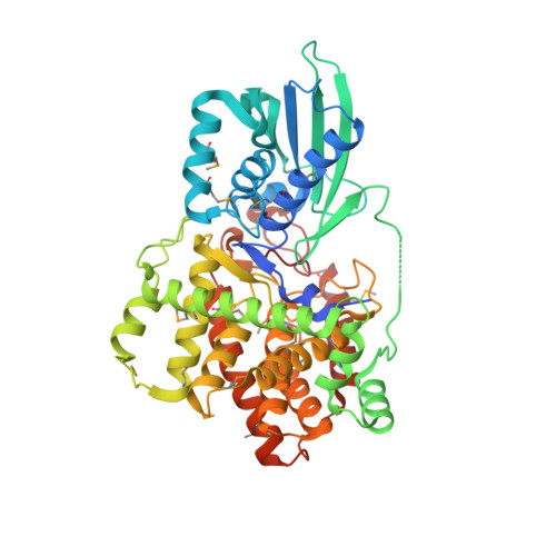
Here we report the first complete structure of a bacterial Fe-S l-serine dehydratase determined to 2.25 Å resolution. The structure is of the type 2 l-serine dehydratase from Legionella pneumophila that consists of a single polypeptide chain containing a catalytic α domain and a β domain that is structurally homologous to the "allosteric substrate binding" or ASB domain of d-3-phosphoglycerate dehydrogenase from Mycobacterium tuberculosis. The enzyme exists as a dimer of identical subunits, with each subunit exhibiting a bilobal architecture. The [4Fe-4S](2+) cluster is bound by residues from the C-terminal α domain and is situated between this domain and the N-terminal β domain. Remarkably, the model reveals that the C-terminal cysteine residue (Cys 458), which is conserved among the type 2 l-serine dehydratases, functions as a fourth ligand to the iron-sulfur cluster producing a "tail in mouth" configuration. The interaction of the sulfhydryl group of Cys 458 with the fourth iron of the cluster appears to mimic the position that the substrate would adopt prior to catalysis. A number of highly conserved or invariant residues found in the β domain are clustered around the iron-sulfur center. Ser 16, Ser 17, Ser 18, and Thr 290 form hydrogen bonds with the carboxylate group of Cys 458 and the carbonyl oxygen of Glu 457, whereas His 19 and His 61 are poised to potentially act as the catalytic base required for proton extraction. Mutation of His 61 produces an inactive enzyme, whereas the H19A protein variant retains substantial activity, suggesting that His 61 serves as the catalytic base. His 124 and Asn 126, found in an HXN sequence, point toward the Fe-S cluster. Mutational studies are consistent with these residues either binding a serine molecule that serves as an activator or functioning as a potential trap for Cys 458 as it moves out of the active site prior to catalysis.
Organizational Affiliation:
Department of Biochemistry, University of Wisconsin , Madison, Wisconsin 53706, United States.