4RJI

Acetolactate synthase from Bacillus subtilis bound to ThDP - crystal form I


Experimental Data Snapshot

  • Method: X-RAY DIFFRACTION
  • Resolution: 3.20 Å
  • R-Value Free: 0.253 
  • R-Value Work: 0.187 
  • R-Value Observed: 0.190 

wwPDB Validation   3D Report Full Report


Ligand Structure Quality Assessment 


This is version 1.3 of the entry. See complete history


Literature

Detailed Structure-Function Correlations of Bacillus subtilis Acetolactate Synthase.

Sommer, B.von Moeller, H.Haack, M.Qoura, F.Langner, C.Bourenkov, G.Garbe, D.Loll, B.Bruck, T.

(2015) Chembiochem 16: 110-118

  • DOI: https://doi.org/10.1002/cbic.201402541
  • Primary Citation of Related Structures:  
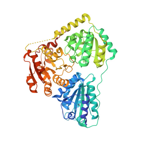
    4RJI, 4RJJ, 4RJK

  • PubMed Abstract: 

    Isobutanol is deemed to be a next-generation biofuel and a renewable platform chemical.1 Non-natural biosynthetic pathways for isobutanol production have been implemented in cell-based and in vitro systems with Bacillus subtilis acetolactate synthase (AlsS) as key biocatalyst.2-6 AlsS catalyzes the condensation of two pyruvate molecules to acetolactate with thiamine diphosphate and Mg(2+) as cofactors. AlsS also catalyzes the conversion of 2-ketoisovalerate into isobutyraldehyde, the immediate precursor of isobutanol. Our phylogenetic analysis suggests that the ALS enzyme family forms a distinct subgroup of ThDP-dependent enzymes. To unravel catalytically relevant structure-function relationships, we solved the AlsS crystal structure at 2.3 Å in the presence of ThDP, Mg(2+) and in a transition state with a 2-lactyl moiety bound to ThDP. We supplemented our structural data by point mutations in the active site to identify catalytically important residues.


  • Organizational Affiliation

    Fachgebiet Industrielle Biokatalyse, Technische Universität München, Lichtenbergstrasse 4, 85748 Garching (Germany).


Macromolecules
Find similar proteins by:  (by identity cutoff)  |  3D Structure
Entity ID: 1
MoleculeChains Sequence LengthOrganismDetailsImage
Acetolactate synthase
A, B, C, D
587Bacillus subtilis PY79Mutation(s): 0 
Gene Names: U712_18080
EC: 4.1.3.18
Entity Groups  
Sequence Clusters30% Identity50% Identity70% Identity90% Identity95% Identity100% Identity
Sequence Annotations
Expand
  • Reference Sequence
Small Molecules
Ligands 3 Unique
IDChains Name / Formula / InChI Key2D Diagram3D Interactions
TPP
Query on TPP

Download Ideal Coordinates CCD File 
E [auth A],
G [auth B],
J [auth C],
L [auth D]
THIAMINE DIPHOSPHATE
C12 H19 N4 O7 P2 S
AYEKOFBPNLCAJY-UHFFFAOYSA-O
PG4
Query on PG4

Download Ideal Coordinates CCD File 
I [auth B]TETRAETHYLENE GLYCOL
C8 H18 O5
UWHCKJMYHZGTIT-UHFFFAOYSA-N
MG
Query on MG

Download Ideal Coordinates CCD File 
F [auth A],
H [auth B],
K [auth C],
M [auth D]
MAGNESIUM ION
Mg
JLVVSXFLKOJNIY-UHFFFAOYSA-N
Experimental Data & Validation

Experimental Data

  • Method: X-RAY DIFFRACTION
  • Resolution: 3.20 Å
  • R-Value Free: 0.253 
  • R-Value Work: 0.187 
  • R-Value Observed: 0.190 
  • Space Group: P 41 21 2
Unit Cell:
Length ( Å )Angle ( ˚ )
a = 140.647α = 90
b = 140.647β = 90
c = 238.877γ = 90
Software Package:
Software NamePurpose
PHENIXrefinement

Structure Validation

View Full Validation Report


Ligand Structure Quality Assessment 


Entry History 

Deposition Data

Revision History  (Full details and data files)

  • Version 1.0: 2014-10-22
    Type: Initial release
  • Version 1.1: 2014-11-26
    Changes: Database references
  • Version 1.2: 2015-01-07
    Changes: Database references
  • Version 1.3: 2023-09-20
    Changes: Data collection, Database references, Derived calculations, Refinement description