4RI3

Crystal structure of DCCD-modified PsbS from spinach


Experimental Data Snapshot

  • Method: X-RAY DIFFRACTION
  • Resolution: 2.70 Å
  • R-Value Free: 0.284 
  • R-Value Work: 0.232 
  • R-Value Observed: 0.237 

wwPDB Validation   3D Report Full Report


Ligand Structure Quality Assessment 


This is version 1.3 of the entry. See complete history


Literature

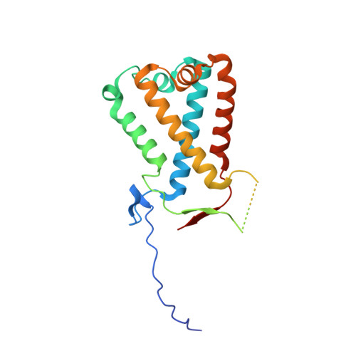
Crystal structures of the PsbS protein essential for photoprotection in plants.

Fan, M.Li, M.Liu, Z.Cao, P.Pan, X.Zhang, H.Zhao, X.Zhang, J.Chang, W.

(2015) Nat Struct Mol Biol 22: 729-735

  • DOI: https://doi.org/10.1038/nsmb.3068
  • Primary Citation of Related Structures:  
    4RI2, 4RI3

  • PubMed Abstract: 

    The photosystem II protein PsbS has an essential role in qE-type nonphotochemical quenching, which protects plants from photodamage under excess light conditions. qE is initiated by activation of PsbS by low pH, but the mechanism of PsbS action remains elusive. Here we report the low-pH crystal structures of PsbS from spinach in its free form and in complex with the qE inhibitor N,N'-dicyclohexylcarbodiimide (DCCD), revealing that PsbS adopts a unique folding pattern, and, unlike other members of the light-harvesting-complex superfamily, it is a noncanonical pigment-binding protein. Structural and biochemical evidence shows that both active and inactive PsbS form homodimers in the thylakoid membranes, and DCCD binding disrupts the lumenal intermolecular hydrogen bonds of the active PsbS dimer. Activation of PsbS by low pH during qE may involve a conformational change associated with altered lumenal intermolecular interactions of the PsbS dimer.


  • Organizational Affiliation

    National Laboratory of Biomacromolecules, Institute of Biophysics, Chinese Academy of Sciences, Beijing, China.


Macromolecules
Find similar proteins by:  (by identity cutoff)  |  3D Structure
Entity ID: 1
MoleculeChains Sequence LengthOrganismDetailsImage
Photosystem II 22 kDa protein, chloroplastic
A, B
212Spinacia oleraceaMutation(s): 0 
Membrane Entity: Yes 
UniProt
Find proteins for Q02060 (Spinacia oleracea)
Explore Q02060 
Go to UniProtKB:  Q02060
Entity Groups  
Sequence Clusters30% Identity50% Identity70% Identity90% Identity95% Identity100% Identity
UniProt GroupQ02060
Sequence Annotations
Expand
  • Reference Sequence
Small Molecules
Ligands 3 Unique
IDChains Name / Formula / InChI Key2D Diagram3D Interactions
BNG
Query on BNG

Download Ideal Coordinates CCD File 
D [auth A],
J [auth B],
K [auth B],
L [auth B]
nonyl beta-D-glucopyranoside
C15 H30 O6
QFAPUKLCALRPLH-UXXRCYHCSA-N
DCW
Query on DCW

Download Ideal Coordinates CCD File 
C [auth A],
I [auth B]
DICYCLOHEXYLUREA
C13 H24 N2 O
ADFXKUOMJKEIND-UHFFFAOYSA-N
HG
Query on HG

Download Ideal Coordinates CCD File 
E [auth A]
F [auth A]
G [auth A]
H [auth A]
M [auth B]
E [auth A],
F [auth A],
G [auth A],
H [auth A],
M [auth B],
N [auth B],
O [auth B]
MERCURY (II) ION
Hg
BQPIGGFYSBELGY-UHFFFAOYSA-N
Experimental Data & Validation

Experimental Data

  • Method: X-RAY DIFFRACTION
  • Resolution: 2.70 Å
  • R-Value Free: 0.284 
  • R-Value Work: 0.232 
  • R-Value Observed: 0.237 
  • Space Group: P 21 21 21
Unit Cell:
Length ( Å )Angle ( ˚ )
a = 71.704α = 90
b = 77.697β = 90
c = 93.159γ = 90
Software Package:
Software NamePurpose
HKL-2000data collection
PHENIXmodel building
PHENIXrefinement
HKL-2000data reduction
HKL-2000data scaling
PHENIXphasing

Structure Validation

View Full Validation Report


Ligand Structure Quality Assessment 


Entry History 

Deposition Data

Revision History  (Full details and data files)

  • Version 1.0: 2015-08-12
    Type: Initial release
  • Version 1.1: 2015-08-26
    Changes: Database references
  • Version 1.2: 2015-09-16
    Changes: Database references
  • Version 1.3: 2020-07-29
    Type: Remediation
    Reason: Carbohydrate remediation
    Changes: Advisory, Data collection, Derived calculations, Structure summary