4RE6

Acylaminoacyl peptidase complexed with a chloromethylketone inhibitor


Experimental Data Snapshot

  • Method: X-RAY DIFFRACTION
  • Resolution: 2.55 Å
  • R-Value Free: 0.260 
  • R-Value Work: 0.212 
  • R-Value Observed: 0.214 

wwPDB Validation   3D Report Full Report


Ligand Structure Quality Assessment 


This is version 1.3 of the entry. See complete history


Literature

Catalytically distinct states captured in a crystal lattice: the substrate-bound and scavenger states of acylaminoacyl peptidase and their implications for functionality.

Menyhard, D.K.Orgovan, Z.Szeltner, Z.Szamosi, I.Harmat, V.

(2015) Acta Crystallogr D Biol Crystallogr 71: 461-472

  • DOI: https://doi.org/10.1107/S1399004714026819
  • Primary Citation of Related Structures:  
    4RE5, 4RE6

  • PubMed Abstract: 

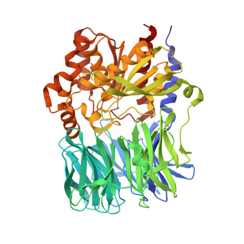
    Acylaminoacyl peptidase (AAP) is an oligopeptidase that only cleaves short peptides or protein segments. In the case of AAP from Aeropyrum pernix (ApAAP), previous studies have led to a model in which the clamshell-like opening and closing of the enzyme provides the means of substrate-size selection. The closed form of the enzyme is catalytically active, while opening deactivates the catalytic triad. The crystallographic results presented here show that the open form of ApAAP is indeed functionally disabled. The obtained crystal structures also reveal that the closed form is penetrable to small ligands: inhibitor added to the pre-formed crystal was able to reach the active site of the rigidified protein, which is only possible through the narrow channel of the propeller domain. Molecular-dynamics simulations investigating the structure of the complexes formed with longer peptide substrates showed that their binding within the large crevice of the closed form of ApAAP leaves the enzyme structure unperturbed; however, their accessing the binding site seems more probable when assisted by opening of the enzyme. Thus, the open form of ApAAP corresponds to a scavenger of possible substrates, the actual cleavage of which only takes place if the enzyme is able to re-close.


  • Organizational Affiliation

    MTA-ELTE Protein Modelling Research Group, Pázmány Péter sétány 1/A, 1117 Budapest, Hungary.


Macromolecules
Find similar proteins by:  (by identity cutoff)  |  3D Structure
Entity ID: 1
MoleculeChains Sequence LengthOrganismDetailsImage
Acylamino-acid-releasing enzyme
A, B, C, D
582Aeropyrum pernix K1Mutation(s): 0 
Gene Names: APE_1547.1
EC: 3.4.19.1
UniProt
Find proteins for Q9YBQ2 (Aeropyrum pernix (strain ATCC 700893 / DSM 11879 / JCM 9820 / NBRC 100138 / K1))
Explore Q9YBQ2 
Go to UniProtKB:  Q9YBQ2
Entity Groups  
Sequence Clusters30% Identity50% Identity70% Identity90% Identity95% Identity100% Identity
UniProt GroupQ9YBQ2
Sequence Annotations
Expand
  • Reference Sequence
Biologically Interesting Molecules (External Reference) 1 Unique
Experimental Data & Validation

Experimental Data

  • Method: X-RAY DIFFRACTION
  • Resolution: 2.55 Å
  • R-Value Free: 0.260 
  • R-Value Work: 0.212 
  • R-Value Observed: 0.214 
  • Space Group: P 1
Unit Cell:
Length ( Å )Angle ( ˚ )
a = 71.58α = 105.15
b = 97.3β = 103.96
c = 99.16γ = 100.26
Software Package:
Software NamePurpose
MxCuBEdata collection
MOLREPphasing
REFMACrefinement
XDSdata reduction
XSCALEdata scaling

Structure Validation

View Full Validation Report


Ligand Structure Quality Assessment 


Entry History 

Deposition Data

Revision History  (Full details and data files)

  • Version 1.0: 2015-01-28
    Type: Initial release
  • Version 1.1: 2015-03-18
    Changes: Database references
  • Version 1.2: 2020-10-21
    Changes: Data collection, Derived calculations
  • Version 1.3: 2023-09-20
    Changes: Data collection, Database references, Refinement description