4R5D

Crystal structure of computational designed leucine rich repeats DLRR_G3 in space group F222


Experimental Data Snapshot

  • Method: X-RAY DIFFRACTION
  • Resolution: 2.53 Å
  • R-Value Free: 0.246 
  • R-Value Work: 0.181 
  • R-Value Observed: 0.185 

wwPDB Validation   3D Report Full Report


This is version 1.4 of the entry. See complete history


Literature

Control of repeat-protein curvature by computational protein design.

Park, K.Shen, B.W.Parmeggiani, F.Huang, P.S.Stoddard, B.L.Baker, D.

(2015) Nat Struct Mol Biol 22: 167-174

  • DOI: https://doi.org/10.1038/nsmb.2938
  • Primary Citation of Related Structures:  
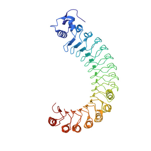
    4R58, 4R5C, 4R5D, 4R6F, 4R6G, 4R6J

  • PubMed Abstract: 

    Shape complementarity is an important component of molecular recognition, and the ability to precisely adjust the shape of a binding scaffold to match a target of interest would greatly facilitate the creation of high-affinity protein reagents and therapeutics. Here we describe a general approach to control the shape of the binding surface on repeat-protein scaffolds and apply it to leucine-rich-repeat proteins. First, self-compatible building-block modules are designed that, when polymerized, generate surfaces with unique but constant curvatures. Second, a set of junction modules that connect the different building blocks are designed. Finally, new proteins with custom-designed shapes are generated by appropriately combining building-block and junction modules. Crystal structures of the designs illustrate the power of the approach in controlling repeat-protein curvature.


  • Organizational Affiliation

    1] Department of Biochemistry, University of Washington, Seattle, Washington, USA. [2] Institute for Protein Design, University of Washington, Seattle, Washington, USA.


Macromolecules
Find similar proteins by:  (by identity cutoff)  |  3D Structure
Entity ID: 1
MoleculeChains Sequence LengthOrganismDetailsImage
Leucine rich repeat protein441synthetic constructMutation(s): 0 
Entity Groups  
Sequence Clusters30% Identity50% Identity70% Identity90% Identity95% Identity100% Identity
Sequence Annotations
Expand
  • Reference Sequence
Experimental Data & Validation

Experimental Data

  • Method: X-RAY DIFFRACTION
  • Resolution: 2.53 Å
  • R-Value Free: 0.246 
  • R-Value Work: 0.181 
  • R-Value Observed: 0.185 
  • Space Group: F 2 2 2
Unit Cell:
Length ( Å )Angle ( ˚ )
a = 91.134α = 90
b = 136.384β = 90
c = 161.753γ = 90
Software Package:
Software NamePurpose
CrystalCleardata collection
PHASERphasing
REFMACrefinement
HKL-2000data reduction
HKL-2000data scaling

Structure Validation

View Full Validation Report


Entry History 

Deposition Data

Revision History  (Full details and data files)

  • Version 1.0: 2015-01-07
    Type: Initial release
  • Version 1.1: 2015-01-14
    Changes: Database references
  • Version 1.2: 2015-01-28
    Changes: Database references
  • Version 1.3: 2015-02-18
    Changes: Database references
  • Version 1.4: 2024-02-28
    Changes: Data collection, Database references, Derived calculations