4R42

Crystal structure of KatB, a manganese catalase from Anabaena PCC7120


Experimental Data Snapshot

  • Method: X-RAY DIFFRACTION
  • Resolution: 1.90 Å
  • R-Value Free: 0.160 
  • R-Value Work: 0.134 
  • R-Value Observed: 0.136 

wwPDB Validation   3D Report Full Report


This is version 1.5 of the entry. See complete history


Literature

KatB, a cyanobacterial Mn-catalase with unique active site configuration: Implications for enzyme function.

Bihani, S.C.Chakravarty, D.Ballal, A.

(2016) Free Radic Biol Med 93: 118-129

  • DOI: https://doi.org/10.1016/j.freeradbiomed.2016.01.022
  • Primary Citation of Related Structures:  
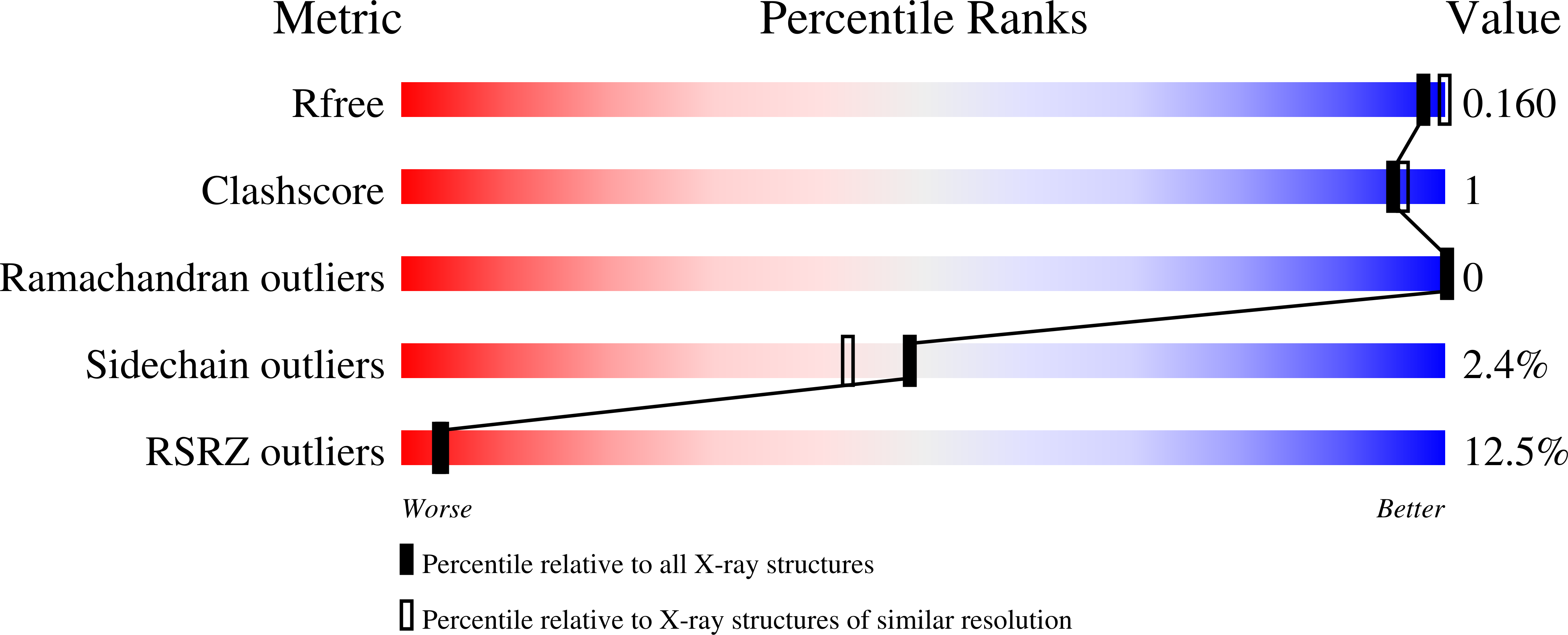
    4R42

  • PubMed Abstract: 

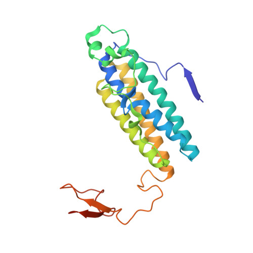
    Manganese catalases (Mn-catalases), a class of H2O2 detoxifying proteins, are structurally and mechanistically distinct from the commonly occurring catalases, which contain heme. Active site of Mn-catalases can serve as template for the synthesis of catalase mimetics for therapeutic intervention in oxidative stress related disorders. However, unlike the heme catalases, structural aspects of Mn-catalases remain inadequately explored. The genome of the ancient cyanobacterium Anabaena PCC7120, shows the presence of two Mn-catalases, KatA and KatB. Here, we report the biochemical and structural characterization of KatB. The KatB protein (with a C-terminal his-tag) was over-expressed in Escherichia coli and purified by affinity chromatography. On the addition of Mn(2+) to the E. coli growth medium, a substantial increase in production of the soluble KatB protein was observed. The purified KatB protein was an efficient catalase, which was relatively insensitive to inhibition by azide. Crystal structure of KatB showed a hexameric assembly with four-helix bundle fold, characteristic of the Ferritin-like superfamily. With canonical Glu4His2 coordination geometry and two terminal water ligands, the KatB active site was distinctly different from that of other Mn-catalases. Interestingly, the KatB active site closely resembled the active sites of ruberythrin/bacterioferritin, bi-iron members of the Ferritin-like superfamily. The KatB crystal structure provided fundamental insights into the evolutionary relationship within the Ferritin-like superfamily and further showed that Mn-catalases can be sub-divided into two groups, each with a distinct active site configuration.


  • Organizational Affiliation

    Solid State Physics Division, Bhabha Atomic Research Centre, Mumbai 400085, India. Electronic address: scbihani@barc.gov.in.


Macromolecules
Find similar proteins by:  (by identity cutoff)  |  3D Structure
Entity ID: 1
MoleculeChains Sequence LengthOrganismDetailsImage
Alr3090 protein
A, B, C
236Nostoc sp. PCC 7120 = FACHB-418Mutation(s): 1 
Gene Names: alr3090
UniProt
Find proteins for Q8YSJ5 (Nostoc sp. (strain PCC 7120 / SAG 25.82 / UTEX 2576))
Explore Q8YSJ5 
Go to UniProtKB:  Q8YSJ5
Entity Groups  
Sequence Clusters30% Identity50% Identity70% Identity90% Identity95% Identity100% Identity
UniProt GroupQ8YSJ5
Sequence Annotations
Expand
  • Reference Sequence
Small Molecules
Ligands 6 Unique
IDChains Name / Formula / InChI Key2D Diagram3D Interactions
P6G
Query on P6G

Download Ideal Coordinates CCD File 
S [auth C]HEXAETHYLENE GLYCOL
C12 H26 O7
IIRDTKBZINWQAW-UHFFFAOYSA-N
PG4
Query on PG4

Download Ideal Coordinates CCD File 
I [auth A]TETRAETHYLENE GLYCOL
C8 H18 O5
UWHCKJMYHZGTIT-UHFFFAOYSA-N
PGE
Query on PGE

Download Ideal Coordinates CCD File 
H [auth A],
R [auth C]
TRIETHYLENE GLYCOL
C6 H14 O4
ZIBGPFATKBEMQZ-UHFFFAOYSA-N
PEG
Query on PEG

Download Ideal Coordinates CCD File 
N [auth B]DI(HYDROXYETHYL)ETHER
C4 H10 O3
MTHSVFCYNBDYFN-UHFFFAOYSA-N
MN
Query on MN

Download Ideal Coordinates CCD File 
D [auth A]
E [auth A]
J [auth B]
K [auth B]
O [auth C]
D [auth A],
E [auth A],
J [auth B],
K [auth B],
O [auth C],
P [auth C]
MANGANESE (II) ION
Mn
WAEMQWOKJMHJLA-UHFFFAOYSA-N
CA
Query on CA

Download Ideal Coordinates CCD File 
F [auth A],
G [auth A],
L [auth B],
M [auth B],
Q [auth C]
CALCIUM ION
Ca
BHPQYMZQTOCNFJ-UHFFFAOYSA-N
Experimental Data & Validation

Experimental Data

  • Method: X-RAY DIFFRACTION
  • Resolution: 1.90 Å
  • R-Value Free: 0.160 
  • R-Value Work: 0.134 
  • R-Value Observed: 0.136 
  • Space Group: P 41 21 2
Unit Cell:
Length ( Å )Angle ( ˚ )
a = 101.404α = 90
b = 101.404β = 90
c = 136.198γ = 90
Software Package:
Software NamePurpose
XSCALEdata scaling
PHENIXrefinement
PDB_EXTRACTdata extraction
MAR345dtbdata collection
XDSdata reduction
XDSdata scaling
PHASERphasing

Structure Validation

View Full Validation Report


Entry History 

Deposition Data

  • Released Date: 2015-08-19 
  • Deposition Author(s): Bihani, S.C.

Revision History  (Full details and data files)

  • Version 1.0: 2015-08-19
    Type: Initial release
  • Version 1.1: 2016-04-06
    Changes: Experimental preparation
  • Version 1.2: 2016-04-20
    Changes: Experimental preparation
  • Version 1.3: 2017-11-22
    Changes: Refinement description
  • Version 1.4: 2019-03-06
    Changes: Data collection, Database references
  • Version 1.5: 2024-03-20
    Changes: Data collection, Database references, Derived calculations