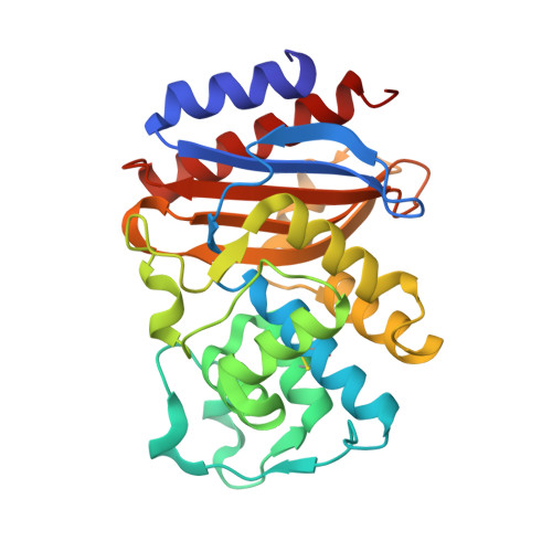
Detecting a Quasi-stable Imine Species on the Reaction Pathway of SHV-1 beta-Lactamase and 6 beta-(Hydroxymethyl)penicillanic Acid Sulfone.
Che, T., Rodkey, E.A., Bethel, C.R., Shanmugam, S., Ding, Z., Pusztai-Carey, M., Nottingham, M., Chai, W., Buynak, J.D., Bonomo, R.A., van den Akker, F., Carey, P.R.(2015) Biochemistry 54: 734-743
- PubMed: 25536850
- DOI: https://doi.org/10.1021/bi501197t
- Primary Citation of Related Structures:
4R3B - PubMed Abstract:
For the class A β-lactamase SHV-1, the kinetic and mechanistic properties of the clinically used inhibitor sulbactam are compared with the sulbactam analog substituted in its 6β position by a CH2OH group (6β-(hydroxymethyl)penicillanic acid). The 6β substitution improves both in vitro and microbiological inhibitory properties of sulbactam. Base hydrolysis of both compounds was studied by Raman and NMR spectroscopies and showed that lactam ring opening is followed by fragmentation of the dioxothiazolidine ring leading to formation of the iminium ion within 3 min. The iminium ion slowly loses a proton and converts to cis-enamine (which is a β-aminoacrylate) in 1 h for sulbactam and in 4 h for 6β-(hydroxymethyl) sulbactam. Rapid mix-rapid freeze Raman spectroscopy was used to follow the reactions between the two sulfones and SHV-1. Within 23 ms, a 10-fold excess of sulbactam was entirely hydrolyzed to give a cis-enamine product. In contrast, the 6β-(hydroxymethyl) sulbactam formed longer-lived acyl-enzyme intermediates that are a mixture of imine and enamines. Single crystal Raman studies, soaking in and washing out unreacted substrates, revealed stable populations of imine and trans-enamine acyl enzymes. The corresponding X-ray crystallographic data are consonant with the Raman data and also reveal the role played by the 6β-hydroxymethyl group in retarding hydrolysis of the acyl enzymes. The 6β-hydroxymethyl group sterically hinders approach of the water molecule as well as restraining the side chain of E166 that facilitates hydrolysis.
Organizational Affiliation:
Department of Biochemistry, ‡Department of Molecular Biology and Microbiology, §Department of Pharmacology, and ∥Department of Medicine, Case Western Reserve University , Cleveland, Ohio 44106, United States.